Emergency War Surgery (2004) - page 7
Abdominal Injuries
ο Even if ductal injury is not identified, it should be
presumed and drained.
ο Resect clearly nonviable pancreatic body/tail tissue.
Transection or near-transection of the pancreatic duct can be
treated by
ο Distal end of proximal pancreas segment oversewn/stapled.
ο Proximal end of distal segment oversewn/stapled and
entire distal segment left in-situ.
ο Distal segment resection (typically requires splenectomy).
ο Distal segment drainage by Roux-en-Y anastomosis to
small bowel.
Major injuries to the head of the pancreas may require
pancreaticoduodenectomy, which SHOULD NEVER BE
ATTEMPTED in an austere environment but instead
treated by the principles of damage control surgery—
DRAIN, DRAIN, DRAIN.
Liver Injuries
Most liver injuries can be successfully treated with direct
pressure and packing followed by aggressive resuscitation
and correction of coagulopathy.
Generous exposure is required and should be gained early
and aggressively.
ο Mobilize triangular and coronary ligaments for full exposure
ο Use extension into right chest if needed.
ο Place several laparotomy pads above the dome of the liver
to displace it down into the field of view.
Short duration clamping of hepatic artery and portal vein (Pringle
maneuver) may be required to slow bleeding while gaining other
control. If bleeding continues despite Pringle maneuver, especially
from behind the liver, this indicates a retrohepatic venous injury
or retrohepatic vena caval injury. The injuries should be
approached in only the most advanced settings with extraordinary
amounts of resources. On table retriage or aggressive packing
and intensive care unit (ICU) resuscitation should be employed.
Use finger fracture of liver parenchyma to expose deep
bleeding vessels.
17.11
Emergency War Surgery
Large exposed injuries of the liver parenchyma can be
controlled in a number of ways:
ο Exposed large vessels and ducts should be suture-ligated.
ο Overlapping mattress sutures of No. 0-Chromic on a blunt liver
needle is fast and effective for controlling raw surface bleeding.
ο Placement of Surgicel on the raw surface and high-power
electrocautery to “weld” it in place is also effective.
Bleeding tracts through the liver can be controlled by tying
off the end of a Penrose drain, placing it through the tract,
and “inflating” it with saline to tamponade the tract.
Urgent surgical resection is strongly discouraged:
ο Indicated only when packing/pressure fails.
ο Follows functional or injury pattern, not anatomic lines.
Use a pedicle of omentum in a large defect to reduce dead space.
Avoidance of coagulopathy, hypothermia and acidosis is
essential in successful management of major liver injuries.
APPLY DAMAGE CONTROL TECHNIQUES EARLY.
Retrohepatic vena cava and
hepatic vein injuries require a
tremendous amount of resources
(blood products, OR time,
equipment) typically unavailable
in a forward surgery setting (on-
table triage in mass casualty).
ο Packing is most successful
option.
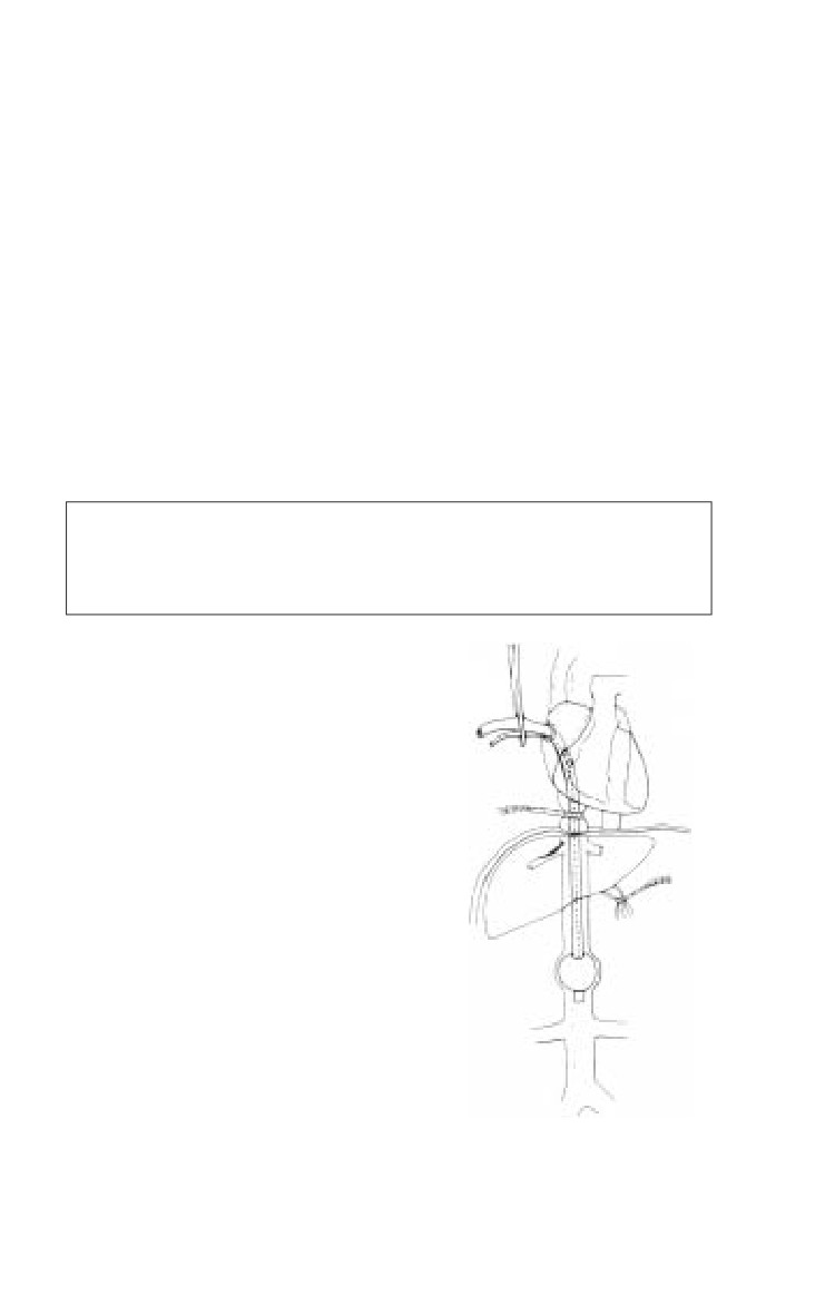
ο If packing fails, consider an atrio-
caval shunt. (see Figure 17-8).
Provide generous closed suction
drainage around major liver injuries.
Biliary Tract Injuries
Injuries to the gall bladder are
treated by cholecystectomy.
Repair common bile duct injuries
over a T-tube.
Fig. 17-8. Atrio-caval shunt.
17.12
Abdominal Injuries
ο A No. 4-0 or smaller absorbable suture is used on the biliary tree.
Extensive segmental loss requires choledochoenterostomy or tube
choledochostomy (depending on time and patient physiology).
Drain widely.
Splenic Injuries
Splenic salvage has no place in combat surgery.
Drains should not be routinely placed postsplenectomy if the
pancreas is uninvolved.
Splenic injury should prompt exploration for associated
diaphragm, stomach, pancreatic, and renal injuries.
Immunize post-op with pneumococcal, haemophilus, and
meningococcal vaccines (may defer until Level 3/CONUS
MTF, but must not be forgotten).
Small-Bowel Injuries
Debride wound edges to freshly bleeding tissue.
Close enterotomies in one or two layers (skin stapler is a rapid
alternative).
With multiple enterotomies to one segment of less than 50%
of small-bowel length, perform single resection with primary
anastomosis. Avoid multiple resections.
Colon Injury
Simple, isolated colon injuries are uncommon. In indigenous
populations and enemy combatants (eg, patients who cannot
be readily evacuated), diversion with colostomy should be
the procedure of choice, especially at Level 2. The often poor
nutritional status of these populations does not support
primary repair. The presence of any of the complicating
factors listed below mandates colostomy.
Simple, isolated colon injuries should be repaired primarily.
ο Debride wound edges to normal, noncontused tissue.
ο Perform two-layer closure or anastomosis.
For complex injuries, strongly consider colostomy/diversion,
especially when associated with:
ο Massive blood transfusion requirement.
ο On-going hypotension.
ο Hypoxia (severe pulmonary injury).
17.13
Emergency War Surgery
ο Reperfusion injury (vascular injury).
ο Multiple other injuries.
ο High-velocity injuries.
ο Extensive local tissue damage.
Potential breakdown of a repair or anastomosis is highest in
the setting of concomitant pancreatic injury.
Damage control technique: control contamination with
ligation/stapling of bowel, delay creation of the stoma to the
definitive reconstruction.
Clearly document treatment for optimal follow-up
throughout Levels of Care.
At the time of formation, a colostomy should be matured.
Rectal Injuries
Rectal injuries can be difficult to diagnose unless very
dramatic. Any question of an injury raised by proximity of
another injury, rectal examination, or plain abdominal film
radiography MANDATES proctoscopy. Gentle distal washout
with dilute Betadine solution is usually required to be able
to perform rigid proctoscopy. Findings can be dramatic
disruptions of the rectal wall but more commonly are subtle
punctuate hemorrhages of the mucosa. All abnormal findings
should prompt corrective intervention.
Consider the traditional 4 “Ds” of rectal injury: Diversion,
Debridement, Distal washout, and Drainage.
ο Of these, diversion is the most important.
♦ Transabdominal sigmoid colostomy is easiest.
♦ If the injury has not violated the peritoneum,
exploration of the extraperitoneal rectum should NOT
be done at laparotomy unless indicated for an associated
nonbowel injury. This avoids contaminating the
abdominal cavity with stool.
ο Debridement and closure of small- to medium-sized
wounds is unnecessary in patients who have been
diverted and drained. In any but the lowest of wounds,
debridement and closure are difficult and troublesome.
ο Distal washout is usually necessary to assess the injury. Use
gentle pressure when irrigating to minimize contamination
of the perirectal space.
17.14
Abdominal Injuries
ο Fecal contamination of the
perirectal space mandates
presacral drainage. Presacral
drains should be placed any
time the patient will leave
your immediate care.
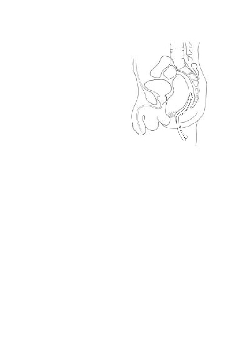
♦ Drains are placed
through the perineum
into the retrorectal space
(Fig. 17-9).
Peritonealized rectal injuries are
easily accessed transabdominally
and should be repaired and
protected with diversion.
Hematoma in the perirectal space
should be drained either
transluminally by leaving the
injury open or by placing
presacral and/or intraabdominal
Fig. 17-9. Presacral drain.
drains.
Retroperitoneal Injuries
Left medial visceral rotation moves the colon, pancreas,
and small bowel to expose the aorta rapidly. Proximal
aortic control can be rapidly obtained with compression
or a clamp on the aorta at the hiatus, or through the left
chest.
Right medial visceral rotation (colon plus Kocher maneuver to
elevate duodenum) exposes the subhepatic vena cava.
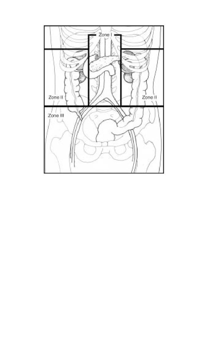
Three zones of the retroperitoneum (Fig. 17-10).
ο I-Central, supracolic: explore for all injuries.
ο II-Central, infracolic: penetrating trauma, explore; blunt
trauma, explore for expanding hematoma.
ο III-Lateral: blunt trauma, avoid exploration if possible
because exploration increases the likelihood of opening a
stable hematoma and, thus, precipitating nephrectomy.
Explore for penetrating trauma.
Gain proximal vascular control before entering the
hematoma.
17.15
Emergency War Surgery
Fig. 17-10. Three zones of the retroperitoreum.
Abdominal Closure
Close fascia if possible.
ο Massive swelling associated with large amounts of blood
loss and resuscitation and large injuries may necessitate
temporary closures (see Chapter 12, Damage Control
Surgery). Otherwise, closure is usually possible.
A few penetrating battlefield wounds are isolated, small, and
without visceral contamination, and it is perhaps safe to close
the skin. Most are not, and these patients will be passed
quickly from one surgeon to the next, so the risk of missed
and catastrophic infection is increased; the skin should not
be closed.
Retention sutures are strongly recommended for the same
reasons.
17.16
Chapter 18
Genitourinary Tract Injuries
Introduction
Genitourinary (GU) injuries constitute approximately 5% of the
total injuries encountered in combat. Their treatment adheres
to established surgical principles of hemostasis, debridement,
and drainage. Proper radiographic evaluation prior to surgery
may replace extensive retroperitoneal exploration at the time
of laparotomy in the diagnosis of serious GU injuries.
GU wounds, aside from injuries of the external genitalia,
are typically associated with serious visceral injury.
Renal Injuries
Most renal injuries, except for those of the renal pedicle, are
not acutely life threatening. Undiagnosed or improperly
treated injuries, however, may cause significant morbidity.
While the vast majority of blunt renal injuries will heal
uneventfully with observation and conservative therapy, a
significant number of renal injuries in combat will come from
penetrating wounds and require exploration.
The evaluation of a suspected renal injury is based on
the type of injury, physical examination, and urinalysis.
Hematuria is usually present in patients with renal trauma,
and gross hematuria in the adult patient is concerning for a
significant injury. The absence of hematuria, however, does
not exclude renal trauma. Renal injury must be suspected in
patients who have sustained significant concurrent injuries
such as multiple rib fractures, vertebral body or transverse
process fractures, crushing injuries of the chest or thorax, or
penetrating injury to the flank, chest, or upper abdomen.
18.1
Emergency War Surgery
Adult patients who present with gross hematuria, micro-
scopic hematuria with shock at any time following the injury,
and significant concurrent injury require further evaluation
of their kidneys. Computed tomography (CT) provides
excellent staging of renal injuries and aids in the decision
whether or not to explore the injured kidney.
In the combat setting, many patients require rapid exploration
before definitive radiographic staging can be completed. An
intraoperative single-shot intravenous pyelogram (IVP) is
useful in their evaluation.
ο Procedure for one-shot IVP:
♦ 2 cc/kg of high-dose contrast is injected in either the
ED or OR setting.
♦ A single standard KUB radiograph is obtained 10
minutes following the contrast injection.
ο While high-osmolality contrast (Renografin, Hypaque, or
Conray) is adequate, low-osmolality contrast (Omnipaque,
Isovue, Optiray) is less likely to generate a reaction and is
less toxic to the kidney.
Major renal injuries usually appear as obscured renal
shadows on IVP.
ο Detailed anatomic information regarding the degree of
renal injury or presence of urinary extravasation should
not be expected on the trauma IVP. Delayed films, however,
may improve detection of urinary extravasation.
ο The study should confirm the presence and function of
the contralateral kidney and may demonstrate congenital
anomalies such as renal ectopia or fusion. Understanding
the function of the contralateral kidney is imperative to
sound intraoperative decision making during exploration
and possible salvage of the injured kidney.
Renal trauma is categorized by the extent of damage to the
kidney.
ο Minor injuries.
♦ Consist of renal contusions or shallow cortical lacerations.
♦ Most common after blunt trauma and usually resolve
safely without renal exploration.
♦ Hydration, antibiotics, and bed rest are the cornerstones
of successful nonoperative management.
18.2
Genitourinary Tract Injuries
ο Major injuries.
♦ Consist of deep cortical lacerations (with or without
urinary extravasation), shattered kidneys, renal vascular
pedicle injuries, or total avulsion of the renal pelvis.
♦ There is an 80% incidence of associated visceral injuries
with major renal trauma. Most cases will require a
laparotomy for evaluation and repair of concurrent
intraperitoneal injuries.
♦ Operative intervention includes debridement of
nonviable renal tissue (partial nephrectomy), closure of
the collecting system, and drainage of the retroperitoneal
area.
♦ Kidney preservation should be considered if at all
possible, although total nephrectomy may be required
for the severely damaged kidney or the unstable patient.
Vascular control of the renal pedicle can be obtained prior
to opening the perirenal fascia when control of
hemorrhage from the kidney requires exploration of the
retroperitoneum.
Operative Technique.
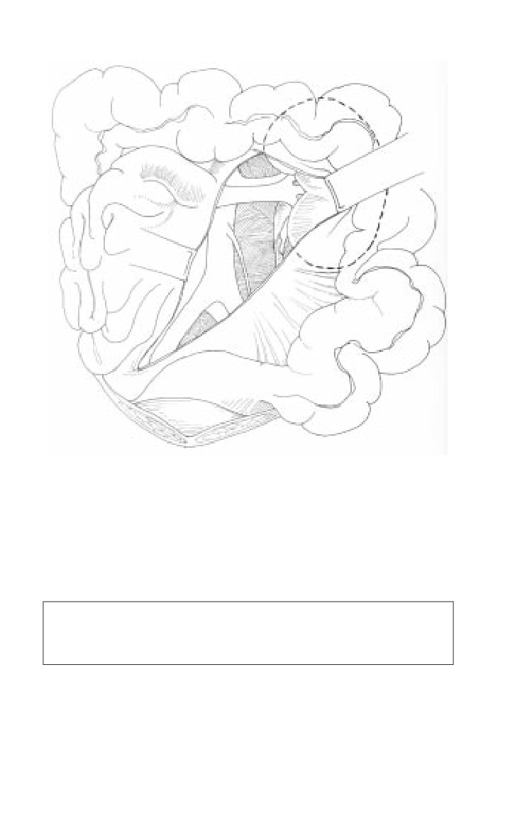
ο Obtain vascular control from a periaortic approach to the
renal vascular pedicle.
♦ The small intestine is retracted laterally and superiorly,
and the posterior peritoneum is incised over the aorta.
♦ The left renal vein, crossing anterior to the aorta, must
be mobilized to gain control of either renal artery.
♦ Atraumatic vascular clamps are used to occlude the
appropriate artery.
ο While vascular control in this fashion may provide the
safest approach against renal hemorrhage and reduce the
likelihood of nephrectomy, it is not a commonly performed
maneuver by either urologists or general surgeons. Direct
reflection of the colon to expose the kidney is feasible (Fig.
18-1). A kidney pedicle clamp should be readily available
for this approach.
18.3
Emergency War Surgery
Fig. 18-1. Exposure of left renal hilum.
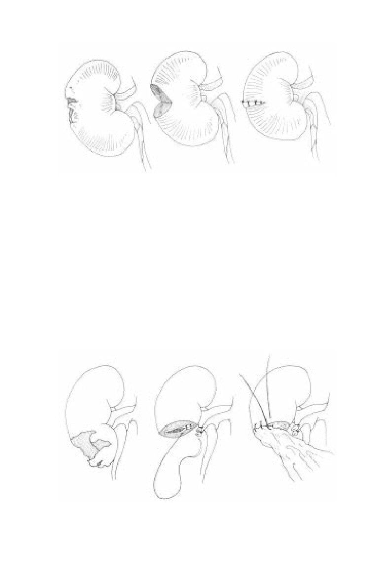
ο Damaged renal parenchyma can be locally debrided (Fig.
18-2), excised in a partial nephrectomy (Fig. 18-3), or
removed in a total nephrectomy depending on the degree
of injury and the condition of the patient.
Nephrectomy may be the best solution for major renal
injuries when associated life-threatening injuries are
present.
ο Watertight closure of the collecting system with absorbable
suture prevents the development of a urine leak.
♦ Urinary diversion is typically unnecessary if formal
renal reconstruction is accomplished.
18.4
Genitourinary Tract Injuries
Fig. 18-2. Steps in renal debridement.
◊ For the sake of expedience or in the presence of
associated injuries of the duodenum, pancreas, or
large bowel, diversion may be required.
◊ Tube nephrostomy, ureteral stent, or ureterostomy
may be utilized.
ο The reconstructed kidney should be covered by perirenal
fat, omentum, or fibrin sealant.
ο A closed suction drain should be left in place.
Fig. 18-3. Steps in partial nephrectomy.
18.5
Emergency War Surgery
Ureteral Injuries
Ureteral injuries are rare but are frequently overlooked
when not appropriately considered. They are more likely
in cases of retroperitoneal hematoma and injuries of the
fixed portions of the colon, duodenum, and spleen.
Isolated ureteral injuries are rare and usually occur in
conjunction with other significant injuries. They can represent
a difficult diagnostic challenge in both the preoperative and
intraoperative settings.
ο Ureteral injuries are not reliably diagnosed by the
preoperative IVP.
ο Hematuria is frequently absent.
ο Blast injury to the urethra may produce significant delayed
complications even when the IVP is normal and the ureter
appears visibly intact. Placement of an indwelling stent is
reasonable when a high-velocity or blast injury occurs in
proximity to the ureter.
ο If a ureteral injury is initially missed and presents in a
delayed fashion, urinary diversion with a nephrostomy
tube and delayed repair at 3-6 months is a safe approach.
Operative Technique.
ο Intraoperative localization of the ureteral injury is
facilitated by IV injection of indigo carmine or direct
injection into the collecting system under pressure.
ο Basic principles of repair.
♦ Minimal debridement.
♦ Primary tension free, 1 cm spatulated anastomosis using
an interrupted single-layer absorbable suture (4-0 or
5-0) closure technique.
♦ Internal (double J ureteral stent) and external drainage.
♦ Lengthening maneuvers.
◊ Ureteral mobilization.
◊ Kidney mobilization.
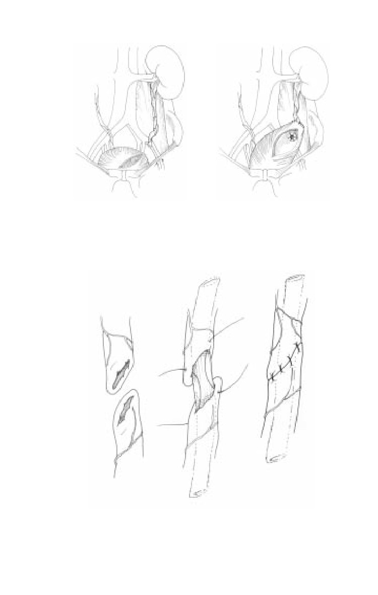
◊ Psoas hitch (Fig. 18-4).
◊ Boare flap.
18.6
Genitourinary Tract Injuries
a
b
Fig. 18-4. The psoas hitch.
Fig. 18-5. Ureteroureterostomy.
18.7

Emergency War Surgery
♦ Isolate repairs with omentum or posterior peritoneum.
ο
The type of repair is based on the following:
♦ Anatomical segment of the traumatized ureter (upper,
middle, and lower third).
♦ Extent of segmental loss.
♦ Other associated injuries.
♦ Clinical stability of the patient.
ο
Upper or Middle ureteral injuries:
♦ Short segment loss/transection: Perform a primary
ureteroureterostomy (Fig. 18-5).
♦ Long segment loss: May require a temporizing tube/
cutaneous ureterostomy with stent placement or
ureteral ligation with tube nephrostomy.
ο
Lower ureteral injuries.
♦ When the injury occurs near the bladder, an ureteroneo-
cystostomy should be performed (Fig. 18-6). This is
typically completed by fixing the bladder to the fascial
covering of the psoas muscle using permanent suture
such as 2.0 or 3.0 Prolene. A transverse cystotomy assists
in elongating the bladder to that location and facilitates
the development of a submucosal tunnel for the
reimplanted ureter.
♦ When a distal ureteral injury is associated with a rectal
injury, ureteral reimplantation is not recommended;
temporary diversion should be performed.
c
a
b
Fig. 18-6. Ureteroneocystostomy.
18.8
Genitourinary Tract Injuries
Ureteral injuries in the combat setting may be best
managed with temporary tube drainage with a small
feeding tube or ureteral stent followed by delayed
reconstruction.
Bladder Injuries
Bladder wounds should be considered in patients with
lower abdominal gunshot wounds, pelvic fractures with
gross hematuria, or those patients unable to void
following abdominal or pelvic trauma.
Bladder disruptions can occur on the intraperitoneal or
extraperitoneal surface of the bladder. The location may
change the symptoms, complications, and management of
this injury.
After ensuring urethral integrity in appropriate cases (see
Urethral Injuries, below), evaluation of the bladder is
performed radiographically with a cystogram.
ο Cystography is performed using a 3-film technique: (1)
scout or plain film KUB concentrating on the pelvis, (2)
full-bladder radiograph after retrograde filling of the
bladder with contrast, and (3) a postdrainage radiograph.
ο Technique: Fill the bladder by gravity with a urethral
catheter using radiopaque contrast medium elevated 20-
30 cm above the level of the abdomen. At least 300 cc (5-7
cc/kg in children) are required for an adequate study. Take
a full-bladder radiograph.
ο Drain the bladder using the catheter and take a postdrainage
radiograph. Small extraperitoneal areas of extravasation
may be apparent only on the postevacuation film.
Operative Technique.
ο Intraperitoneal Injuries.
♦ Cystography reveals contrast medium interspersed
between loops of bowel.
♦ Management consists of immediate exploration,
multilayer repair of the injury with absorbable suture,
suprapubic tube cystostomy, and drainage of the
18.9
Emergency War Surgery
perivesical extraperitoneal space.
ο
Extraperitoneal injuries.
♦ Bladder laceration is most often the result of laceration
by bony fragments from a pelvic fracture.
♦ Cystography reveals a dense, flame-like extravasation
of contrast medium in the pelvis on the postevacuation
film.
♦ The bladder usually heals with 10-14 days of Foley
catheter drainage without the need for primary repair.
If the urine is clear, catheter drainage alone is preferred
for treatment of most extraperitoneal ruptures.
♦ In cases of abdominal exploration for other injuries,
primary repair and drainage are necessary if the extra-
peritoneal space is entered. Repair can be completed
from inside the bladder through a cystotomy to avoid
disturbing any pelvic hematoma. Patients with
concurrent rectal injuries should be managed more
aggressively and may benefit from hematoma evacuation
and primary bladder repair.
Urethral Injuries
A urethral injury should be suspected in patients with a
scrotal hematoma, blood at the meatus, or a floating/high-
riding prostate. Catheterization is contraindicated until
urethral integrity is confirmed by retrograde urethro-
graphy.
Retrograde urethrography is performed to evaluate the
anatomy of the urethra.
ο Take oblique radiographs of the pelvis to avoid “end-on”
imaging that obscures the bulbar urethra.
ο Insert the end of a sterile catheter tip syringe (60 cc) into
the urethral meatus while grasping the glans to prevent
leakage. Alternately, insert an unlubricated Foley catheter
into the fossa navicularis (approximately 3 cm) and inflate
the balloon with 3 cc of water.
ο Gently instill 15-20 cc of water-soluble contrast. The
radiograph is taken during injection.
18.10
Genitourinary Tract Injuries
ο Contrast must be seen flowing into the bladder to clear
the proximal urethra of injury. Posterior urethral injuries
seen in pelvic fractures may be missed otherwise.
ο If no injury is identified, carefully place a Foley catheter.
If any difficulty in passing the catheter is encountered,
the urethra should not be instrumented and a suprapubic
tube cystostomy is performed.
Operative Technique.
ο
The urethra is divided into anterior and posterior
(prostatic) segments by the urogenital diaphragm.
♦ Anterior urethral injuries may result from blunt trauma,
such as results from falls when astride an object
(straddle) or from penetrating injuries.
◊
Blunt trauma resulting in minor nondisruptive
urethral injuries may be managed by gentle insertion
of a 16 French Foley catheter for 7-10 days.
◊
Penetrating wounds should be managed by explora-
tion and judicious debridement.
Small, clean lacerations may be repaired primarily
by reapproximation of the urethral edges using
interrupted 4-0 chromic suture.
Do not mobilize the entire urethra for a primary
anastomosis, because the shortened urethral
length in the pendulous urethra may produce
ventral chordee and an anastomosis under tension.
Instead, marsupialize the injured urethral segment
by suturing the skin edges to the cut edges of the
urethra. Marsupialization should be performed until
healthy urethra is encountered both proximally
and distally. Closure of the marsupialized urethra
is subsequently performed at 6 months to
reestablish urethral continuity.
♦ Posterior urethral disruption commonly occurs
following pelvic fracture injuries.
◊ Rectal examination reveals the prostate to have been
avulsed at the apex.
18.11
Emergency War Surgery
◊ Improved continence and potency rates are attained
when suprapubic tube cystostomy is used as the
initial management.
◊ Suprapubic urinary diversion is maintained for 10-14
days and urethral integrity is confirmed radiograph-
ically prior to removal of the suprapubic tube.
◊ With expectant observation, virtually all these
injuries will heal with an obliterative prostatomem-
branous urethral stricture, which can be repaired
secondarily in 3-6 months after reabsorption of the
pelvic hematoma.
◊ Initial exploration of the pelvic hematoma is strictly
reserved for patients with concomitant bladder neck
or rectal injury.
External Genitalia Injuries
The management of wounds to the penis, scrotum, testes,
or spermatic cord should be as conservative as possible
and consists of hemorrhage control, debridement, and
early repair to prevent deformity.
Injuries to the penis that disrupt Buck’s fascia should be
sutured to prevent further bleeding and avoid future penile
curvature with erection. When extensive penile skin is lost,
the penis may be placed in a scrotal tunnel until a plastic
repair can be performed.
The scrotum is highly vascularized, and extensive debridement
is usually not necessary for scrotal wounds.
ο Most penetrating scrotal injuries should be explored to
evaluate the testicle for injury and reduce the risk of
hematoma formation.
ο Most partial scrotal avulsions are best treated by primary
closure with absorbable 3-0 sutures in two layers.
ο Primary closure is selected for patients without associated
life-threatening injuries who sustained injury less than 8
hours prior. A Penrose drain or small closed drain can be
placed to reduce hematoma formation. The testes can be
18.12
Genitourinary Tract Injuries
placed in protective pockets in the medial thigh for
complete scrotal avulsion.
It is essential, when dealing with testicular wounds, to
conserve as much tissue as possible.
ο Herniated parenchymal tissues should be debrided, and
the tunica albuginea closed by mattress sutures.
ο The testicle is placed in the scrotum or in a protective
pocket in the medial thigh.
ο A testicle should never be resected unless it is hopelessly
damaged and its blood supply destroyed.
18.13
Chapter 19
Gynecologic Trauma and Emergencies
Introduction
The current active duty population consists of 14% women,
many of whom are subject to the same risks of combat injury as
their male colleagues. This chapter deals with OB/GYN
emergencies that may present to a deployed medical treatment
facility (MTF), particularly in military operations other than war
(MOOTW).
Gynecologic Trauma
Vulva
Vulvar injuries include lacerations and hematomas.
ο Lacerations that are superficial, clean, and less than 6 hours
old can be primarily closed with absorbable suture.
Debridement of obviously devitalized tissue is
recommended.
♦ Deep lacerations should be examined and explored to
rule out urethral, anal, rectal mucosa, or periclitoral
injuries.
♦ Placing a urethral catheter will assist in determining
injury. If found, single-layer closure with fine (4-0 or
smaller), absorbable suture, leaving the catheter in place,
is recommended. Rectal and periclitoral injuries are
closed in a similar fashion.
♦ Anal lacerations should be repaired by approximating
the cut ends of the anal sphincter with size 0 or 1
absorbable suture.
♦ Antibiotics (2nd generation cephalosporin) are
recommended with contaminated wounds.
19.1
Emergency War Surgery
Vulvar trauma may cause infrafascial (below the pelvic
diaphragm) hematoma.
ο Because the deeper layer of subcutaneous vulvar fascia is
not attached anteriorly to the pubic rami, hematoma can
spread freely into the anterior abdominal wall.
ο Most vulvar hematomas are treated conservatively.
ο External compression and ice packs should be applied
until hemostasis is ensured by serial examination of the
vulva, vagina, and rectum.
ο Signs of shock in association with a decreasing
hematocrit should prompt consideration of extraperi-
toneal expansion. Ultrasound or computed tomography
is useful for detecting expansion not diagnosed by clinical
exam.
A vulvar hematoma continuing to expand despite
external pressure, or presenting acutely with a size
greater than 10 cm, should be incised and evacuated, with
ligation of bleeding vessels and packing placed to secure
hemostasis.
Vagina
Trauma to the vagina can cause lacerations, and less
commonly, suprafascial (above the pelvic diaphragm)
hematoma.
Vaginal trauma has been reported in approximately 3.5% of
women with traumatic pelvic fractures. Concomitant
urologic trauma, most often involving the bladder and/or
urethra, has been described in about 30% of patients with
vaginal trauma.
Thorough inspection and palpation of the vagina and
rectovaginal exam are necessary for detection of vaginal
trauma and to determine the need for further urologic
evaluation/imaging. Due to pelvic instability (in fracture
cases) or pain, examination under sedation or anesthesia
may be necessary.
Patients with vaginal lacerations typically present with
bleeding, sometimes profusely, from the well-vascularized
vagina.
19.2
Gynecologic Trauma and Emergencies
Lacerations are repaired using the guidelines given above
for vulvar lacerations.
Vaginal hematoma is usually accompanied by severe rectal
pressure and is diagnosed by palpation of a firm, tender mass
bulging into the lateral vagina. Vaginal hematoma should
be treated by incision, evacuation, ligation, and packing.
Unrecognized vaginal trauma can result in dyspareunia,
pelvic abscess, and fistula formation.
Uterus/Cervix
Trauma to the uterus and cervix is most commonly found in
association with pregnancy, but may be seen as a result of
penetrating vaginal or abdominal trauma.
Noninfected simple cervical lacerations should be repaired
to optimize restoration of normal anatomy (and possibly
decrease the risk of cervical incompetence or stenosis with
dysmenorrhea from poor healing). Absorbable 0 grade suture
can be used.
Acute penetrating trauma involving the uterine fundus
usually causes little bleeding and can be managed expectantly
without repair. Damage to the uterine wall with bleeding can
be repaired with size 0 absorbable suture.
Trauma involving the lateral wall of the uterus may cause
significant bleeding, but can usually be controlled by
successive ligation of the ascending and descending branches
of the uterine artery as described below in the obstetrical
section “uterine atony.”
Hemorrhage not responding to ligation, or extensive
mutilating damage to the cervix or uterus, is best treated
by hysterectomy.
Prophylactic antibiotics should be given. Adnexa should be
retained unless there is an indication for removal (see next
page).
19.3
Emergency War Surgery
Basic steps for performing an emergent total
abdominal hysterectomy.
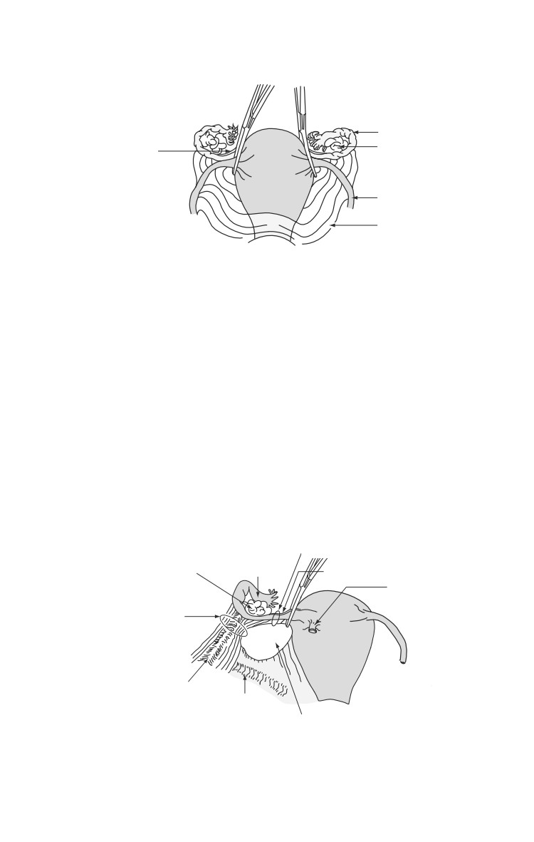
Ligate/cauterize round ligaments (Fig. 19-1).
Incise anterior leaves of broad ligaments bilaterally, then
continue across midline to incise vesicouterine fold.
Mobilize bladder downward by blunt dissection (and
sharp dissection if necessary) from lower uterine
segment and cervix.*
To retain adnexa, clamp/cut/ligate utero-ovarian
ligaments and fallopian tubes near their connections to
uterine fundus (Fig. 19-2).
To remove adnexa with uterus, clamp/cut/ligate
infundibulopelvic ligaments after making windows in
posterior leaves of broad ligaments above ureters.
Incise posterior peritoneum to mobilize adnexa either
away from (if being retained) or toward (if being
removed) uterus.
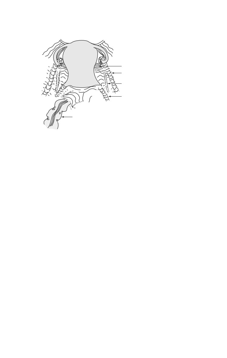
Incise peritoneum overlying rectovaginal space, then
mobilize rectum downward and away from posterior
vagina by blunt dissection (Fig. 19-3).*
Clamp/cut/ligate uterine arteries along lateral surface
of uterus at uterocervical junction, staying within 1 cm
of uterus to avoid damaging ureters.
Clamp/cut/ ligate remainder of cardinal ligaments,
paracervical tissue, and uterosacral ligaments by taking
successive inferior bites until cervicovaginal junction
is reached; each bite should be placed medial to previous
bite to avoid injuring ureter and bladder.
Crossclamp vagina below cervix.
Transect vagina, removing uterus (and attached adnexa,
if applicable).
Suture vaginal cuff closed, ensuring bladder is not
incorporated.
*In case of dense adhesions between cervix and bladder or rectum in
emergent setting, or ongoing hemorrhage with poor visualization,
supracervical hysterectomy can be performed. After mobilizing bladder
and rectum from uterus and ligating uterine arteries, uterine fundus is
transected from cervix with a knife. Cervix is then oversewn with a baseball
stitch, staying medial to ligated uterine arteries.
19.4
Gynecologic Trauma and Emergencies
Anterior View
Fallopian Tube
Utero-Ovarian
Ovary
Ligament
Round Ligament
X
Broad Ligament
Bladder
X: Location to Incise
Peritoneum and Enter Vesicocervical
Uterine Space
Fig. 19-1. Abdominal hysterectomy anterior view.
Adnexa
Fallopian Tubes.
ο Damage to the wall of the fallopian tube by ruptured
ectopic pregnancy or penetrating abdominal trauma
should be treated by salpingectomy if there is significant
damage to the tube, due to the risk of subsequent or recur-
rent ectopic pregnancy if left in situ. If the damage is
Adnexal View
Location for Ligation if Adnexum Retained
Ovary
Fallopian Tube
Utero-Ovarian Ligament
Round
Ligament,
Location for Ligation
Ligated
if Adnexum
Removed
Infundibulopelvic
Ligament
Ureter
Window in Posterior Leaf of Broad Ligament
Fig. 19-2. Abdominal hysterectomy adnexal view.
19.5
Emergency War Surgery
Posterior View
Uterus
Cardinal Ligament
Infundibulopelvic
Ligament
Ureter
X
Uterosacral Ligament
X: Location to Incise Peritoneum
and Enter Rectovaginal Space
Rectosigmoid Colon
Fig. 19-3. Abdominal hysterectomy posterior view.
equivalent to a linear salpingotomy (see below), achieve
hemostasis, then allow healing by secondary intention.
ο The mesosalpinx is ligated or cauterized, then the tube
ligated and cut at its connection with the uterine fundus.
ο Unruptured ampullary/isthmic ectopic pregnancy can be
treated by linear salpingotomy, with extraction of the
ectopic gestation. The tubal incision is left open to heal by
secondary intention.
ο An unruptured or ruptured corneal/interstitial ectopic
pregnancy requires wedge resection of the uterine
cornuum with salpingectomy.
ο An ectopic pregnancy spontaneously aborted into the
abdominal cavity through the end of the tube should be
removed, but the tube may be left in situ if hemostasis is
attained.
Ovaries.
ο A ruptured ovarian cyst should be treated via cystectomy
by shelling the cyst wall out of the ovary, then cauterizing
or ligating any bleeding vessels, usually at the base of the
cyst.
19.6
Gynecologic Trauma and Emergencies
ο Torsion of an ovarian mass is first treated by assessing
the ovary. Untwist the ovary and or fallopian tube. If it
appears healthy with some continuing blood supply, it can
be left in situ. If the ovary contains a large (> 4 cm) simple
appearing cyst, the cyst can be drained and the cyst wall
removed. Interrupted sutures using a fine monofilament
or electrocautery can be used to obtain hemostasis. If the
ovary appears dark and dusky after untwisting, perform
a salpingo-oophorectomy by ligating the infundib-
ulopelvic ligament first (after identifying the ureter), then
the utero-ovarian ligament and fallopian tube.
ο Hemorrhage from an infundibulopelvic ligament, as a
result of penetrating abdominal trauma, is best treated by
ligation with salpingo-oophorectomy.
Retroperitoneal Hematoma
Laceration of an arterial branch of the hypogastric artery
can cause a retroperitoneal hematoma.
A large amount of blood may collect in the broad ligament
with few symptoms. Dissection of the hematoma can extend
up to the level of the renal vessels. The hematoma may be
discovered during emergency surgery for trauma or during
re-operation or post pelvic surgery, or can be prompted by
signs of shock suggesting internal bleeding.
Retroperitoneal hematoma can be treated by hypogastric
artery ligation on the affected side. Bilateral hypogastric
artery ligation may be necessary for hemostasis. The uterus,
tubes and ovaries may be left in situ if viable and without
other indication for removal.
Gynecologic/Obstetric Emergencies
Acute Vaginal Hemorrhage Unrelated to Trauma.
ο Bright red vaginal bleeding filling more than one large
perineal pad per hour is considered vaginal hemorrhage.
A pregnancy test and pelvic exam direct initial therapy.
♦ If the patient is not pregnant, hormonal management
with 25 mg IV Premarin or 50 mcg estrogen-containing
oral birth control pills (OCPs) should be given every 6
hours.
19.7
Emergency War Surgery
◊ If the bleeding responds to hormonal management,
OCPs should be continued qid for 5-7 days while
more definitive diagnosis and management plans are
made.
◊ If the bleeding has not decreased significantly within
24 hours, dilatation and curettage is reasonable. If
the heavy bleeding continues, imaging studies and
possibly coagulation studies will be needed to help
direct further therapy.
♦ In the pregnant patient, heavy bleeding from the
cervical os with uterine size < 20 weeks (fundus at/or
below the level of patient’s umbilicus) suggests
spontaneous abortion. Dilatation and suction curettage
should be performed.
◊
Ectopic pregnancy uncommonly presents with acute
hemorrhage, but should be considered if the patient
has an acute abdomen or if scant tissue is obtained
on curettage.
◊
In a pregnant patient with uterine size consistent with
a third trimester gestation (> 4 cm above the
umbilicus in a singleton pregnancy), vaginal
hemorrhage is usually an indication of placental
abruption or placenta previa.
◊
Emergent cesarean section will be necessary if the
uterine hemorrhage does not spontaneously resolve
within several minutes.
◊
After delivery of the fetus and placenta, persistent
hemorrhage unresponsive to more conservative
measures may require hysterectomy (see emergent
cesarean and uterine atony below).
◊
Pregnant patients (mothers) with acute vaginal
hemorrhage who have Rh negative bloodtype, or if
their Rh status is unknown, should be given
RhoGAM 300 mcg IM.
ο
A hemorrhaging mass in the vagina is most likely cervical
cancer. The vagina should be packed to tamponade the
bleeding after placing a urethral catheter. Placing sutures
is generally futile and may make the bleeding worse.
19.8
Gynecologic Trauma and Emergencies
Precipitous Vaginal Delivery
Preparation.
ο Supplies needed for the delivery, include povidone-iodine
sponges, a 10 cc syringe, lidocaine, 2 Kelly clamps, ring
forceps, dry towels, a bulb syringe, and scissors.
ο The mother should be placed on her left side for labor.
ο The fetal heart rate should be determined every 15 minutes
prior to pushing, and following each contraction during
the pushing phase using a vascular Doppler. Normal heart
rate is between 120-160 bpm. The heart rate often drops
with the contraction, but should recover to normal prior
to the next contraction.
If the heart rate drops below 100 and stays low for more
than 2 minutes, a cesarean section should be considered.
ο When the patient presents, the cervix should be examined
to determine dilation and fetal position. For the woman to
begin pushing, the cervix should be completely dilated
(10 cm) and no cervix should be felt on either side of the
fetal head. If the baby’s head is not presenting, move to
cesarean section immediately. If there is any question, and
ultrasound is available, it should be used to determine the
presentation.
Delivery.
ο Once the patient begins pushing, flex the hips to optimally
open the pelvis. The patient may be on her back, or tilted
slightly to the left. Assistants should support the legs
during pushing and relax them between contractions.
ο Clean the perineum with sterile Betadine solution. If this
is the patient’s first delivery, the perineum should be
anesthetized with lidocaine in case an episiotomy is
needed. There is little support for prophylactic episiotomy,
but may be necessary if the fetus is large, or tearing is
anticipated.
ο The fetal head delivers by extension. Pushing upward on
the fetal chin through the perineum can assist this process.
19.9
Emergency War Surgery
Additionally, it is extremely important to control the rate
of delivery of the head with the opposite hand.
ο
If an episiotomy is needed, it should be cut in the posterior
midline from the vaginal opening approximately 1/2 the
length of the perineum, and extend about 2-3 cm into the
vagina.
ο
After delivery of the head, the mouth and nose should be
suctioned and the neck palpated for evidence of a nuchal
cord. If present, this should be reduced by looping it over
the fetal head, or by clamping twice and cutting if it will
not reduce.
ο
Next, the operator’s hands are placed along the parietal
bones and the patient is asked to push again to allow
delivery of the anterior shoulder. Gentle downward
traction should allow the shoulder to clear the pubis, and
the fetus should be directed anteriorly to allow delivery
of the posterior shoulder. The remainder of the body will
normally follow rapidly. Wrap infant in dry towels.
ο
Once the fetus delivers, the cord should be doubly clamped
and cut. The placenta usually delivers within 15 minutes
of delivery, but may take up to 60 minutes. Delivery of the
placenta is heralded by uterine fundal elevation,
lengthening of the cord, and a gush of blood. While
waiting, gentle pressure may be placed on the cord,
however, vigorous uterine massage and excessive traction
can lead to complications.
ο
Following delivery of the placenta, the patient should be
started on an infusion of lactated Ringer’s with 20 units of
oxytocin (Pitocin). Oxytocin can also be given IM if there
is no IV access. If there is no oxytocin available, alternatives
are methylergonovine maleate (Methergine) 0.2 mg
intramuscular (IM) or allowing the patient to breastfeed.
The placenta should be inspected for evidence of
fragmentation that can indicate retained products of
conception.
Inspection and repair.
ο Following delivery of the placenta, the vagina and cervix
should be inspected for lacerations. Downward digital
pressure on the posterior vagina and fundal pressure (by
19.10
Gynecologic Trauma and Emergencies
an assistant, if available) will facilitate visualization of the
cervix. A ring forceps is then used to grasp and visualize
the entire cervix.
ο
The vagina should be inspected with special attention to
the posterior fornix. The perineum and periurethral areas
should also be inspected. Vaginal and cervical lacerations
may be repaired with 3-0 vicryl or an equivalent suture in
running or interrupted layers.
ο
If the anal sphincter is lacerated, it should be reapprox-
imated with 2-0 absorbable interrupted single or figure-
of-eight sutures.
ο
If the patient has torn into the rectum, the rectal-vaginal
septum should be repaired with interrupted sutures of 3-
0 vicryl. A second layer imbricating the underlying tissue
will decrease the risk of breakdown. Care should be taken
to preserve aseptic technique. If a large tear is noted, a
saddle block or spinal anesthetic may be necessary.
ο
Patients with a periurethral tear may require urethral
catheterization. In addition to lacerations, hematoma in
the vulva, vagina, or retroperitoneum may occur. See above
gynecologic trauma for management.
Emergency Cesarean Section
Indications.
ο Fetal heart rate drops below 100 and stays down for more
than 2 minutes.
ο Acute uterine hemorrhage persisting for more than a few
minutes (suggestive of placental abruption or previa).
ο Breech or transverse fetal presentation.
The patient should be placed in the left tilt position with an
IV bag or towel displacing the uterus to the left. She should
undergo a quick prep from just below the breasts to the mid
thigh. A major abdominal equipment set should have most
of the instruments that you will need.
Basic steps to performing an emergency C-section (Fig. 19-
4 a,b,c,d).
ο Enter the abdomen through lower midline.
19.11
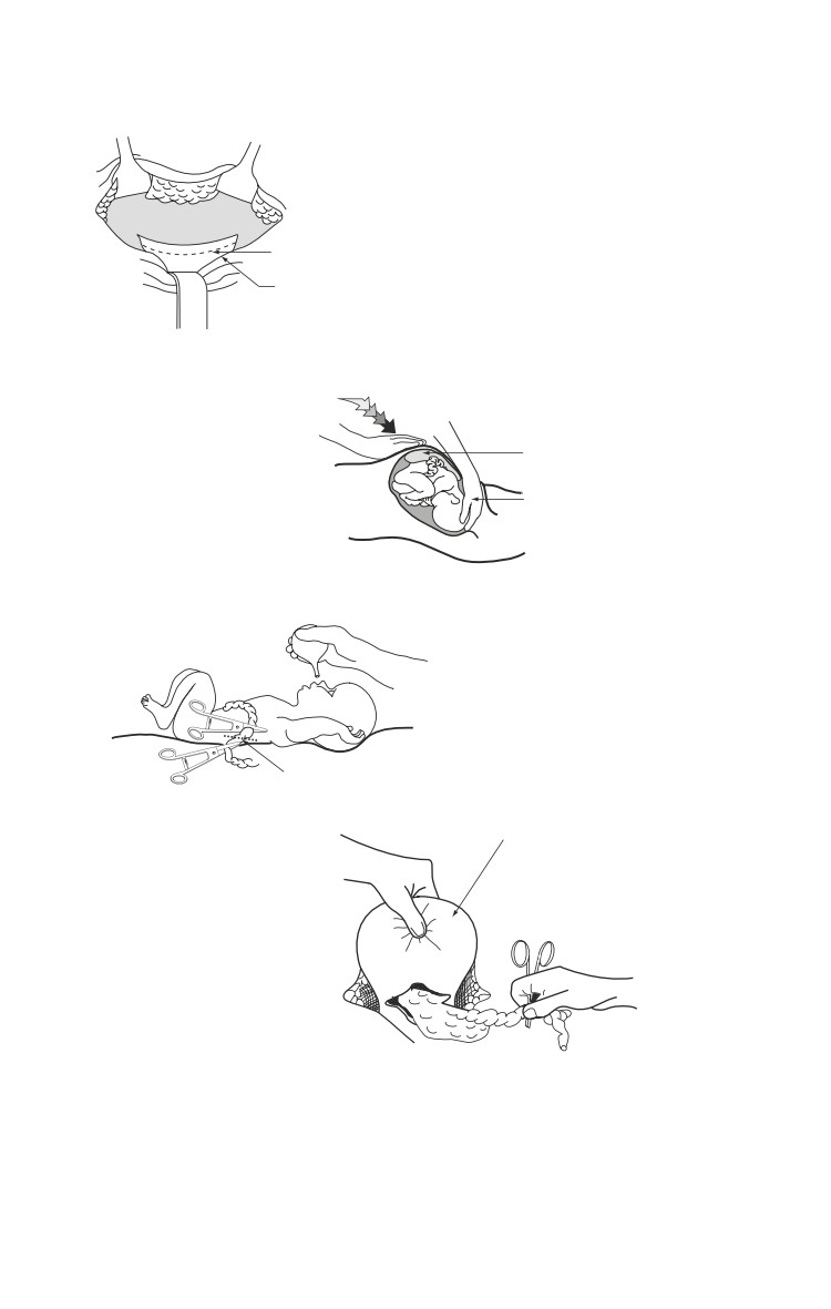
Emergency War Surgery
Uterine Incision
a
Uterus
Incision in Lower
Uterine Segment
Bladder Retracted After
Incising Vesicouterine Fold
b
Delivery of Fetus
Fundal Pressure
Exerted
Placenta
Operator's Hand
in Position to
Deliver Fetal Head
c
Delivered Infant on Abdomen
Nose and Mouth Suctioned
Umbilical Cord Doubly Clamped,
Then Cut
Uterine Fundus Exteriorized
d
Fig. 19-4. Emergency C-Section.
19.12
Gynecologic Trauma and Emergencies
ο
Identify and incise the peritoneal reflection of the bladder
transversely, and create a bladder flap to retract the bladder
out of the field.
ο
Using a scalpel, carefully incise the uterus transversely
across the lower uterine segment (where the uterine wall
thins).
ο
Once the amniotic membranes are visible or opened,
extend the incision laterally, either bluntly or by carefully
using bandage scissors. Avoid the uterine vessels laterally.
If necessary, the incision can be extended at one or both of
its lateral margins in a J-fashion by vertical incision.
ο
Elevate the presenting fetal part into the incision, with an
assistant providing fundal pressure.
ο
Upon delivery of the fetus, suction the nose and mouth
and clamp and cut the cord. Hand the infant off for care
(see below).
ο
Direct anesthetist to administer 2 grams of cefazolin
(Ancef) once the cord is clamped.
ο
Allow the placenta to deliver by providing gentle traction
on the cord and performing uterine massage.
ο
Begin oxytocin, if available as above.
ο
Using a sponge, clean the inside of the uterus, and
vigorously massage the fundus to help the uterus contract.
ο
Quickly close the incision with 0-vicryl. A single layer
(running, locking) is adequate, if hemostatic, for transverse
incisions. Take care to avoid the lateral vessels. If the
incision has a vertical extension, close it in 2 or 3 layers.
ο
Once hemostasis is assured, close the fascia and abdomen
in the usual fashion.
ο
In the rare case of continued uterine hemorrhage, evaluate
and treat as outlined in the section below.
Uterine Atony
The majority of postpartum hemorrhage is secondary to
uterine atony (failure of uterine contracture).
When the uterus fails to contract following delivery of
the placenta, bleeding may be torrential and fatal.
19.13
Emergency War Surgery
Initial management should include manual uterine explor-
ation for retained placenta. Without anesthesia, this procedure
is painful. An opened sponge is placed around the examiner’s
fingers. Place the opposite hand on the patient’s uterine
fundus and apply downward pressure. Gently guide your
fingers through the open cervix and palpate for retained
placenta. The inside of the uterus should feel smooth, and
retained placenta will feel like a soft mass of tissue. This may
be removed manually or by using a large curette if available.
If no tissue is encountered, use both hands to apply vigorous
uterine massage to improve the uterine tone.
Medications should also be used if available. Oxytocin may
be given by IV bolus using 40 units in 1000 cc, or up to 10
units IM, but never by IV push. Although unlikely to be
available, other medications that can be considered are
Methergine, dinoprostone (Prostin), and misoprostol
(Cytotec).
If no medication is available, the patient should be
encouraged to breast feed or do nipple stimulation to increase
endogenous oxytocin release.
If conservative measures fail to arrest the postpartum
hemorrhage, laparotomy (if the hemorrhage is occurring
post vaginal delivery), should be performed.
Intraoperative massage of the uterine fundus may be tried.
If the massage fails to improve uterine tone, the uterine
arteries should be ligated in a stepwise fashion. Begin with
the ascending branch at the junction of the upper and lower
uterine segment. Using 0 or No. 1 chromic, place a stitch
through the myometrium medial to the artery from front to
back. The stitch is then brought out through the adjacent
broad ligament and tied. If bilateral ligation of the ascending
branch does not control bleeding, the descending branch
should be ligated at the level of the uterosacral ligament. If
this fails, consider bilateral hypogastric artery ligation (see
above). If this fails, proceed to hysterectomy as outlined in
the gynecologic portion of this chapter.
19.14
Gynecologic Trauma and Emergencies
Neonatal Resuscitation
Immediately following delivery, every infant should be
assessed for need for resuscitation. Equipment that may be
needed includes warm towels, bulb syringe, stethoscope,
flow-inflating or self-inflating bag with oxygen source,
laryngoscope and blade, suction catheter, and endotracheal
tube. The two medications that may be needed are epine-
phrine 1:10,000 and naloxone (Narcan) 0.4 mg/ml.
Nearly 90% of term babies are delivered without risk factors
and with clear fluid, requiring only to be dried, suctioned
and observed. If the baby is less than 36 weeks, or if there
is meconium in the fluid at delivery, the baby will need to
be observed more closely.
ο
In the first 30 seconds after delivery, dry and stimulate the
baby, position it in order to open the airway, and give free
flow oxygen if the color is poor.
ο
At 30 seconds, evaluate the heart rate. If it is < 100 begin
to provide positive pressure ventilation. After 30 seconds
of ventilation, recheck the heart rate. If it is < 60, then
chest compressions should be started. After 30 seconds
of chest compressions, again re-evaluate. If the heart rate
remains < 60 you should administer epinephrine.
Epinephrine can be given either through the umbilical vein
or the endotracheal tube. The level of experience of the
team present should dictate which route should be used.
The dose is 0.1-0.3 ml/kg of the 1:10,000 solution.
ο
If heart rate rises over 100, stop the positive pressure
ventilations, but continue to provide free flow oxygen. If
the mother has been given a dose of narcotics in the 4 hours
prior to delivery, and positive pressure ventilation has
resulted in a normal heart rate and color but poor
respiratory effort, then naloxene is indicated. Administer
naloxene by IV, IM, or endotracheal route at a dosage of
0.1 mg/kg.
If at any time during resuscitation the heart rate goes above
100, with good respiratory effort, tone and color, the baby
may be moved to an observation status.
19.15
Chapter 20
Wounds and Injuries of the Spinal
Column and Cord
Introduction
Combat injuries of the spinal column, with or without associated
spinal cord injury, differ from those encountered in civilian
practice. These injuries are often open, contaminated, and
usually associated with other organ injuries.
Following the ABCs of advanced trauma life support (ATLS),
management principles include:
Initial spine stabilization to prevent neurologic deterioration.
Diagnosis.
Definitive spinal stabilization.
Functional recovery.
In complete injuries, the likelihood of neurological
recovery is minimal and is not influenced by emergent
surgical intervention. However, incomplete injuries with
neurological deterioration may benefit from emergent
surgical decompression. Emergent, life-saving, soft
tissue exploration, and debridement may still be
required, particularly with colorectal involvement.
Classification
Four discriminators must be considered in the classification and
treatment of spinal injuries.
Is injury open or closed?
Neurologic status: complete vs incomplete vs intact.
ο Complete injury demonstrates no neurologic function
below the level of injury after the period of spinal shock
20.1
Emergency War Surgery
(usually 24-48 h, evidenced by return of the bulbocavern-
osus reflex).
Location of the injury: cervical, thoracic, lumbar, or sacral.
Degree of bony and ligamentous disruption: stable vs
unstable.
Pathophysiology of the Injury to the Spinal Cord
Injury to the spinal cord is the result of both primary and
secondary mechanisms.
ο Primary: the initial mechanical injury due to local
deformation and energy transmission.
♦ High-velocity missile wounds in the paravertebral area
can cause injuries even without direct trauma.
Stretching of the tissue around the missile’s path during
formation of the temporary cavity, or fragmentation of
the projectile and bone resulting in secondary missiles,
cause injury without any direct destruction of the spinal
column.
The destructive nature of high-velocity wounds explains
the futility of decompressive laminectomy in the
management of these wounds.
ο Secondary: the cascade of biochemical and cellular
processes initiated by the primary process that causes
cellular damage and even cell death.
The critical care of spinal cord injury patients includes
attempts to minimize secondary injury from hypoxia,
hypotension, hyperthermia, and edema.
Mechanical integrity of the vertebral column
The vertebral column is composed of three structural columns
(Table 20-1).
20.2
Wounds and Injuries of the Spinal Column and Cord
Table 20-1. Support of the Spinal Column.
Column Bony Elements
Soft-Tissue Elements
Anterior Anterior two-thirds of vertebral Anterior longitudinal ligament
body
Anterior annulus fibrosus
Middle
Posterior one-third of vertebral Posterior longitudinal ligament
body
Posterior annulus fibrosus
Pedicles
Posterior Lamina
Ligamentum flavum
Spinous processes
Interspinous ligaments
Facet joints
Injuries occur by either direct penetrating forces or a
combination of flexion, axial loading, rotation, and distraction
forces.
Loss of integrity of two of the three columns results in
instability of the spine.
Instability is common following blunt injury of the
vertebral column, but is not usually the case with gunshot
or fragment wounds of the vertebral column.
Cervical instability by lateral radiograph (must include C-
7/T-1 junction) is defined by:
ο 3.5 mm or greater sagittal displacement or translation.
ο Angulation of 11° or more on the lateral view.
ο Should questions exist regarding cervical stability, flexion
and extension lateral radiographs can be obtained in the
awake, cooperative patient.
Thoracic and lumbar spine instability:
ο 5 mm of sagittal translation.
ο 20°-30° of sagittal angulation.
ο 50% loss of vertebral body height.
ο Widened pedicles on anterior-posterior (AP) radiographs.
Computed tomography (CT) is very effective in demonstrating
spinal instability and has become available in some field
environments.
20.3
Emergency War Surgery
Instability must be presumed (and the spine stabilized) in
any patient with:
Complaints of a sense of instability (holds his head
in his hands).
Vertebral column pain.
Tenderness in the midline over the spinous
processes.
Neurologic deficit.
Altered mental status.
SUSPECTED, but NOT PROVEN injury.
Patient Transport
On the battlefield, preservation of the life of the casualty
and medic are of paramount importance. In these circum-
stances, EVACUATION TO A MORE SECURE AREA
TAKES PRECEDENCE OVER SPINE IMMOBILIZA-
TION. Data do not support the use of cervical collars and
spine boards for PENETRATING spine injuries on the
battlefield.
Extrication
Cervical spine.
ο The neck should never be hyperextended.
ο If an airway is needed.
♦ If appropriate, attempt endotracheal intubation with in-
line neck stabilization.
♦ Cricothyroidotomy is necessary if intubation fails.
ο The head should be maintained in alignment with the
body.
♦ Requires several people, including one just to stabilize
the neck.
♦ Log roll with the most experienced person stabilizing
the neck.
ο A stiff cervical collar and sandbags provide stabilization
of the neck during the transport. The head and body should
be secured to the extrication device.
Thoracic and lumbar spine.
20.4
Wounds and Injuries of the Spinal Column and Cord
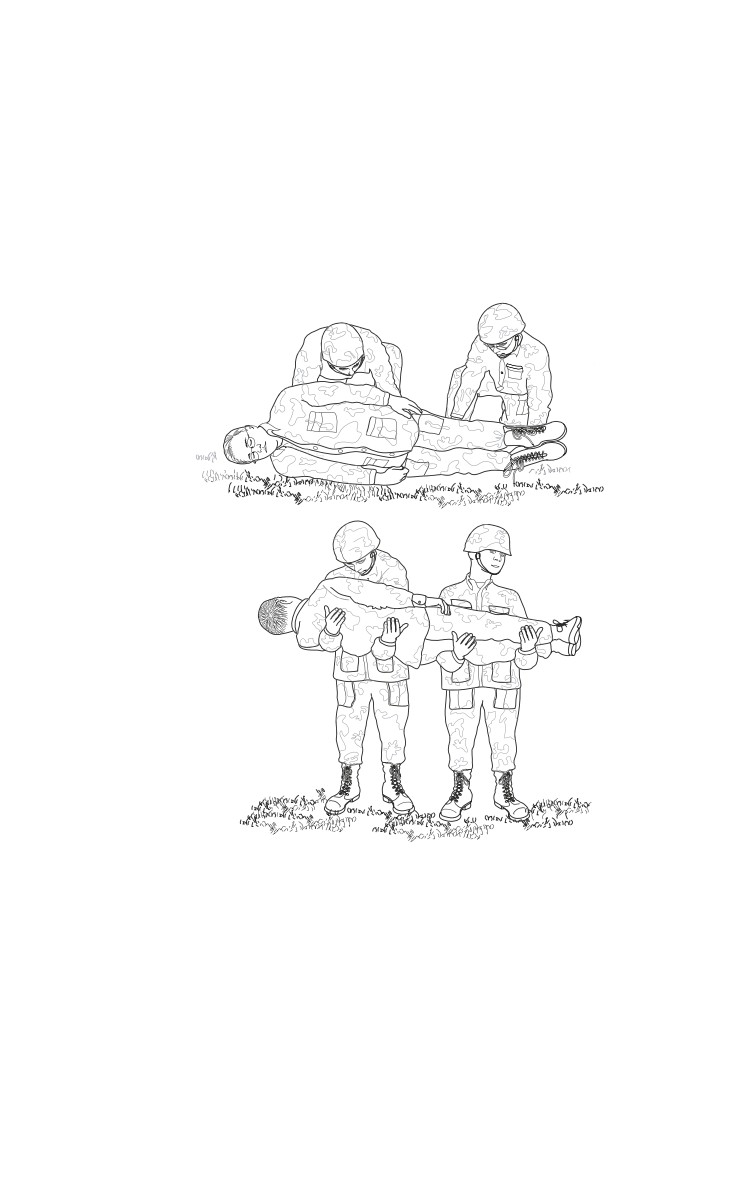
ο Use log roll or two-man carry as demonstrated in Fig. 20-1.
♦ The two-man carry alone does not protect the cervical
spine.
♦ The cradle-drop drag may also be used.
ο In the absence of a spine board, makeshift litters can be
fashioned from local materials.
a
b
Fig. 20-1. (a) Log roll (b) two-man carry.
Anatomical Considerations
Cervical Spine
All potentially unstable cervical spine injuries should be immob-
ilized in a rigid collar, unless halo immobilization is required.
Indications for halo use:
20.5