Emergency War Surgery (2004) - page 6
Emergency War Surgery
Blast over-pressure CNS injuries.
ο A force transmitted by the great vessels of the chest to the
brain; associated with unconsciousness, confusion,
headache, tinnitus, dizziness, tremors, increased startle
response, and occasionally (in the most severe forms)
increased ICP. Bleeding may occur from multiple orifices
including ears, nose, and mouth.
A combination of multiple injury types are typically involved
in combat-related brain injuries. Those injuries generally involve
the face, neck, and orbit; entry wounds may be through the
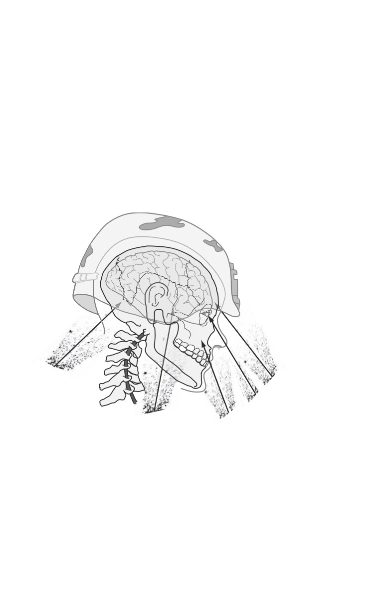
upper neck, face, orbit, or temple (Fig. 15-1).
Trans-suboccipital
Transfrontal
Transorbital
Transtemporal
Transfacial/infratemporal
Fig. 15-1. Common vectors of penetrating injury.
The subocciput, occiput, and retroauricular regions are
overlooked most. Injuries to these areas can indicate underlying
injury to the posterior fossa, major venous sinus, or carotid
artery, as fragments pass through the skull base. Reconstructing
the fragment path based on combination of plain films and
15.2
Head Injuries
computed tomography (CT) can be challenging. In transorbital,
transtemple, or penetrating injuries that cross the midline, an
underlying injury to intracranial vessels should be suspected with
associated pseudoaneurysms, dissections, or venous sinus injury.
Explosion results in flying fragments, with possible vehicular-
collision-associated blunt injuries. Depending on the proximity
to the explosion, a blast over-pressure phenomenon may also
result. In a severely brain-injured patient, more deficits than
indicated by the CT scan may be due to possible underlying
injury to brachiocephalic vessels, shear injury, or the effects of
blast over-pressure with resulting cerebral vasospasm. Plain
films, more useful in penetrating than blunt trauma, may reveal
a burst fracture of the skull indicating the tremendous
perforating force of a penetrating missile. Transventricular
bihemispheric fragment tracts portend a poor prognosis.
Severe head injuries are often seen in combination with
significant chest, abdomen, and extremity injuries. Very rapid
hemorrhage control is the priority in the noncranial injuries;
utilizing damage control concepts and focusing attention on the
head injury. All efforts should be directed toward early diagnosis
and intervention of the head injury.
Traditional Classification of Head Injuries
Open injuries are the most commonly encountered brain
injuries in combat.
Closed injuries, seen more often in civilian settings, may have
a higher frequency in military operations other than war.
Scalp injuries may be closed (eg, contusion) or open (eg,
puncture, laceration, or avulsion).
ο Any scalp injury may be associated with a skull fracture
and/or underlying brain injury.
ο Open scalp injuries bleed profusely, even to the point of lethal
blood loss, but usually heal well when properly repaired.
Skull fractures may be open or closed, and are described as
linear, comminuted, or depressed.
ο Skull fractures are usually associated with some degree of
brain injury, varying from mild concussion, to devastating
diffuse brain injury, to intracranial hematomas.
15.3
Emergency War Surgery
ο Open skull fractures are prone to infection if not properly
treated.
Mechanisms of Injury
Primary injury is a function of the energy transmitted to the
brain by the offending agent.
ο Very little can be done by healthcare providers to influence
the primary injury.
ο Enforcement of personal protective measures (eg, helmet,
seatbelts) by the command is essential prevention.
Secondary injury results from disturbance of brain and
systemic physiology by the traumatic event.
Hypotension and hypoxia are the two most acute and easily
treatable mechanisms of secondary injury.
ο Other etiologies include seizures (seen in 30%-40% of patients
with penetrating brain injuries), fever, electrolyte disturbances
(specifically, hyponatremia or hyperglycemia), and infection.
ο All of the above conditions can be treated.
ο Elevations of ICP may occur early as a result of a space-
occupying hematoma, or develop gradually as a result of
brain edema or hydrocephalus.
ο Normal ICP is 5-15 mm Hg, with normal cerebral
perfusion pressure (CPP = MAP-ICP) usually around 70-
80 mm Hg.
ο Decreases in perfusion pressure as a result of systemic
hypotension or elevated ICP gradually result in alteration
of brain function
(manifested by impairment of
consciousness), and may progress to global brain ischemia
and death if not treated.
Patient Assessment and Triage
During the primary and secondary assessment, attention
should be placed on a complete examination of the scalp and
neck. Fragments that enter the cranial vault with a
transtemple, transorbital, or cross midline trajectory should
be suspected as having associated neurovascular injuries.
Wounds are typically contaminated by hair, dirt, and debris
15.4
Head Injuries
and should be copiously irrigated clean with control of scalp
hemorrhage but not at the expense of delaying definitive
neurosurgical treatment! Scalp hemorrhage can be controlled
with a head wrap, scalp clips, or surgical staples; a meticulous
plastic surgical closure is only appropriate after intracranial
injuries have been ruled out.
The most important assessment is the vital signs.
Next is the level of consciousness, best measured and
recorded by the Glasgow Coma Scale (GCS) (see below).
GLASGOW COMA SCALE
Component
Response
Score
Motor Response
Obeys verbal command
6
(best extremity)
Localizes pain
5
Flexion-withdrawal
4
Flexion (decortication)
3
Extension (decerebration)
2
No response (flaccid)
1
Subtotal
(1-6)
Eye Opening
Spontaneously
4
To verbal command
3
To pain
2
None
1
Subtotal
(1-4)
Best Verbal Response
Oriented and converses
5
Disoriented and converses
4
Inappropriate words
3
Incomprehensible sounds
2
No verbal response
1
Subtotal
(1-5)
Total
(3-15)
Triage decisions in the patient with craniocerebral trauma
should be made based on admission GCS score.
ο A GCS < 5 indicates a dismal prognosis despite aggressive
comprehensive treatment and the casualty should be
considered expectant.
15.5
Emergency War Surgery
ο A GCS > 8 indicates that a casualty may do well if managed
appropriately.
♦ In general, neurologically stable patients with
penetrating head injury can be managed effectively in
the ICU with airway and ventilatory support,
antibiotics, and anticonvulsants while awaiting surgery.
♦ An exception to this would be a deteriorating patient
with a large hematoma seen on CT—this should be
considered a surgical emergency.
ο Casualties with GCS 6-8 can be the most reversible, with
forward neurosurgical management involving control of
ICP and preservation of CBF.
Another important assessment is pupillary reactivity.
A single dilated or nonreactive pupil adds urgency and
implies the presence of a unilateral space-occupying lesion
with secondary brain shift. Immediate surgery is indicated.
ο The presence of bilateral dilated or nonreactive pupils is a
dismal prognostic sign in the setting of profound alteration
of consciousness.
Radiographic evaluation.
ο Deployable CT scanners in standard ISO shelters are
increasingly available in the field environment. To keep
the scanner operational, a qualified maintenance chief
should be married to the scanner (“crew-chief” concept).
♦ CT is the definitive radiographic study in the evaluation
of head injury, and should be employed liberally as it
greatly improves diagnostic accuracy and facilitates
management.
ο Skull radiographs still have a place in the evaluation of
head injury (especially penetrating trauma).
♦ In the absence of CT capability, AP and lateral skull
radiographs help to localize foreign bodies in cases of
penetrating injuries and can also demonstrate skull
fractures.
♦ This can help direct otherwise “blind” surgical
intervention initially to the side of the head where the
fracture is identified.
15.6
Head Injuries
ο Cervical spine injury is uncommon in the setting of
penetrating head injury.
♦ Closed head injury is commonly associated with injury
of the cervical spine.
♦ Assume the presence of cervical spine injury and keep
the cervical spine immobilized with a rigid collar until
standard AP, lateral, and open-mouth radiographs can
be obtained to exclude injury.
♦ CT once again is useful in evaluating casualties with a
high suspicion for spinal injury.
Management
Medical.
ο
Primary tenets are basic but vital; clear the airway,
ensure adequate ventilation, and assess and treat for
shock (excessive fluid administration should be
avoided).
ο
In general, patients with a GCS < 12 should be managed
in the ICU.
ο
ICU management should be directed at the avoidance
and treatment of secondary brain injury.
♦ PaO2 should be kept at a minimum of 100 mm Hg.
♦ PCO2 maintained between 35 and 40 mm Hg.
♦ The head should be elevated approximately 30°.
♦ Sedate patient and/or pharmacologically paralyze to
avoid “bucking” the ventilator and causing ICP
spikes.
♦ Broad-spectrum antibiotics should be administered
to patients with penetrating injuries (a third-
generation cephalosporin, vancomycin or Ancef,
Unasyn or meropenen if acinetobacter suspected).
♦ Anaerobic coverage with metronidazole should be
considered for grossly contaminated wounds or those
whose treatment has been delayed more than 18 hours.
♦ Phenytoin should be administered in a 17-mg/kg
load, which may be placed in a normal saline
piggyback and given over 20-30 minutes (no more
than 50 mg/min, because rapid infusion may cause
cardiac conduction disturbances).
15.7
Emergency War Surgery
◊ A maintenance dose of 300-400 mg/d, either in
divided doses or once before bedtime, should be
adequate to maintain a serum level of 10-20 µg/L.
♦ Measure serum chemistries daily to monitor for
hyponatremia.
♦ Monitor and treat coagulopathy aggressively.
♦ Monitoring of ICP is recommended for patients with
GCS< 8 (in essence, it is a substitute for a neurologic
examination).
◊
A simple fluid-path monitor usually works well and
allows CSF drainage. It may then be coupled to a
manometer or to a multifunction cardiac monitor
similar to a central venous catheter or arterial line.
Administer prophylactic antibiotic.
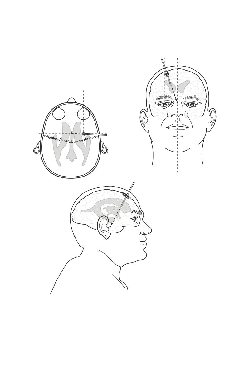
Make an incision just at or anterior to the coronal
suture, approximately 2.5-3 cm lateral to the
midline (Fig. 15-2a,b).
A twist drill craniostomy is performed, the
underlying dura is nicked, and a ventricular
catheter placed into the frontal horn of the
lateral ventricle (encountered at a depth of 5 to
6 cm) (see Fig. 15-2b,c). Catheter should be directed
toward the medial epicanthis on the coronal plane,
and the tragus in the sagittal plane.
Even small ventricles can be easily cannulated
by aiming the tip of the catheter toward the
nasion in the coronal plane.
Ventricular catheters are highly preferable;
acceptable substitutes are an 8 F Robinson
catheter or pediatric feeding tube.
A key feature of this technique is to tunnel the
drain out through a separate incision 2-3 cm
from the primary one, thus reducing the risk of
infection.
◊
The goal of management is to maintain a CPP of 60-
90 mm Hg.
◊
A sustained ICP > 20 mm Hg should be treated (Fig. 15-3).
15.8
Head Injuries
Aim for nasion or
b
ipsilateral medial
epicanthus
a
Midpupiliary
line
Tragus line
Aim for tragus
c
Fig. 15-2. Placement of intracranial ventricular catheter.
15.9

Emergency War Surgery
Levels of Intervention
Pentobarbital
or Thiopental Load
Hemicraniectomy/Duraplasty
Mild Hypothermia (34°-36°C) should be
considered in isolated head
injury but should be avoided in multi-trauma
Mannitol/Lasix (possible 3.0% Hypertonic Saline)
CSF Drainage via Ventriculostomy
Moderate Head-Up Posture (30°)
Adequate Sedation/Analgesia (Versed, Fentanyl, Morphine, Propofol)
Fig. 15-3. Levels of intervention to reduce ICP.
Sedation, head elevation, and paralysis.
CSF drainage if a ventricular catheter is in place.
Hyperventilation to a PCO2 of 30 to 35 mm Hg only
until other measures take effect. (Prolonged
levels below this are deleterious as a result of small
vessel constriction and ischemia.)
Refractory intracranial hypertension should be
managed with an initial bolus of 1g/kg of
mannitol and intermittent dosing of 0.25-0.5 g/
kg q4h as needed.
Aggressive treatment with mannitol should be
accompanied by placement of a CVP line or even
a PA catheter because hypovolemia may ensue.
Any patient who develops intracranial hypertension
or deteriorates clinically should undergo prompt
repeat CT.
◊ Mild hypothermia may be considered in isolated
head injury, but avoid in the multitrauma patient.
♦ Treat hypovolemia with albumin, normal saline,
hypertonic saline, or other volume expanders to create
a euvolemic, hyperosmolar patient (290-315 mOsm/L).
♦ Blast over-pressure CNS injuries.
15.10
Head Injuries
◊ Supportive medical therapy is usually sufficient.
Only in rare cases is an ICP monitor, ventriculostomy,
or cranial decompression necessary. In the absence
of hematomas the use of magnesium has been
beneficial. Structures particularly sensitive include
optic apparatus, hippocampus, and basal ganglia.
Delayed intracranial hemorrhages have been
reported. Additionally, these patients have a higher
susceptibility to subsequent injury and should be
evaluated at a level 4/5 facility. Repetitive injury and
exposure to blast over-pressure may result in
irreversible cognitive deficits.
Surgical
ο
Goals: prevent infection and relieve/prevent intracranial
hypertension.
ο
Indications for emergent exploration.
♦ Space-occupying lesions with neurological changes (eg,
acute subdural/epidural hematoma, abscess).
♦ Intracranial hematoma producing a > 5 mm midline
shift or similar depression of cortex.
♦ Compound depressed fracture with neurological changes.
♦ Penetrating injuries with neurological deterioration.
ο
Relief of ICP with hemicraniectomy/duraplasty/
ventriculostomy.
♦ A large trauma flap should be planned for the
evacuation of a mass lesion with significant underlying
edema in the supratentorial space.
♦ The flap should extend a minimum of 4 cm posterior to
the external auditory canal and 3-4 cm off midline.
Exposing the frontal, temporal, and parietal lobes allows
for adequate cerebral swelling and avoids brain
herniation at the craniotomy edge.
♦ A capacious duraplasty should be constructed with
a subdural ICP/ventricular catheter in place,
allowing monitoring and drainage from the injured
hemisphere.
ο
Shave hair widely and scrub and paint the scalp with
betadine.
ο
General anesthesia for major cases.
15.11
Emergency War Surgery
ο Administer empiric antibiotics
(third-generation
cephalosporin).
ο Positioning can be adequately managed with the head in
a doughnut or horseshoe-type head holder. For unusual
positioning of the head, such as to gain access to the
subocciput, use a standard three-point Mayfield fixation
device.
ο Make a generous scalp incision to create an adequate flap.
♦ The flap should have an adequate pedicle to avoid
ischemia.
♦ Retraction of the scalp flap over a rolled laparotomy
sponge will avoid kinking the flap, which also may lead
to ischemia.
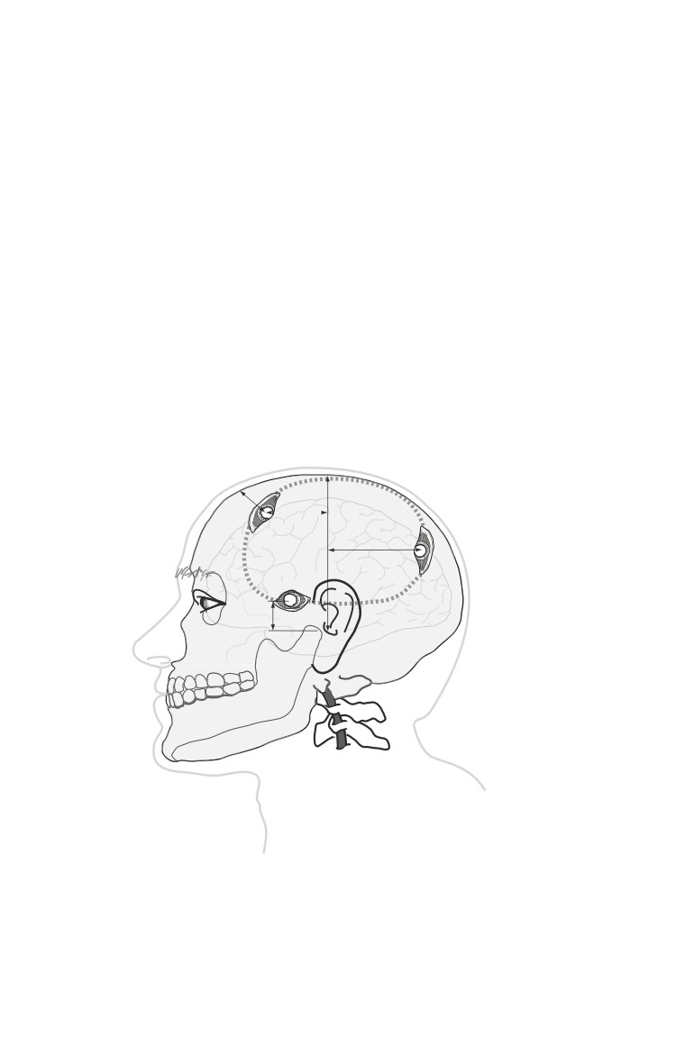
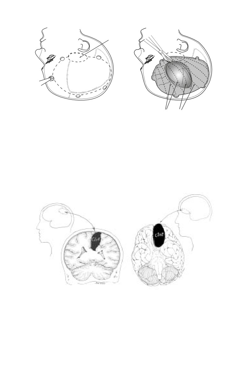
ο The skull should be entered through a series of burr holes
(Fig. 15-4) that are then joined to create a craniotomy flap
(Fig. 15-5a).
2 cm
4–5 cm
5-6 cm
1
cm
zygoma
Fig. 15-4. Cranial landmarks and location of standard burr holes.
15.12
Head Injuries
a
Small temporal
b
craniotomy
Dashed lines
(dural incision sites)
Fig. 15-5. Craniotomy flap and exposed hematoma.
♦ Burr holes alone are inadequate to treat acute
hematomas, but are of diagnostic utility in the absence
of CT scanner. Exploratory burr holes may miss
subfrontal or interhemispheric hematomas (Fig. 15-6).
Fig. 15-6. Hematomas missed with routine exploratory burr holes.
15.13
Emergency War Surgery
♦ The bone work may be done with a Hudson brace
and Gigli’s saw, though a power craniotome is certainly
preferable if available (see Fig. 15-5a).
ο
A dural opening, using the entire expanse of the cranial
opening (with enough edge left to close the dura at the
end of the case), should be created.
♦ The base of the dural opening should be on the side near
any neighboring major venous sinus to avoid injury to
large draining veins and aggravation of cerebral edema.
ο
The hematoma should then be gently evacuated with a
combination of suction, irrigation, and mechanical removal
(see Fig. 15-5b).
ο
Meticulous hemostasis should be achieved and the dura
closed.
ο
Approach to penetrating injury with neurologic changes
is aimed at removal of devitalized brain and easily
accessible foreign bodies.
♦ Perform copious irrigation with an antibiotic solution
(such as bacitracin) and a concerted attempt made to
achieve watertight dural closure (again, using
pericranium, among others, as needed).
♦ Tension-free scalp closure is also essential, but
replacement of multiple skull fragments in an attempt
to reconstruct the skull defect is not appropriate in the
battlefield setting.
◊ Excellent results can be achieved with cranioplasty
after evacuation out of the theater and a sufficient
delay to minimize risk of infection.
ο
If a duraplasty is required, pericranium, temporalis fascia,
or tensor fascia lata may be used.
ο
Tack-up sutures should be placed around the periphery
and in the center of the dural exposure to close the dead
space and discourage post-operative epidural hematoma
formation.
ο
Replace bone flap and secure with wire or heavy suture.
♦ If severe brain swelling precludes replacement of the
bone flap it can be discarded or preserved in an
abdominal-wall pocket.
15.14
Head Injuries
ο The galea of the scalp should generally be closed separately
with an absorbable suture, and with staples used to close
the skin.
♦ A single layer closure with heavy monofilament nylon
is acceptable but should definitely include the galea,
with the sutures remaining in place at least 10 days.
♦ A subgaleal or epidural drain may be used at the
discretion of the surgeon.
ο Apply a snug dressing using roller bandages around the
entire head.
Evacuation of the Severely Head-Injured Patient
The trip is always longer than advertised. Transport only
patients who can be expected to survive 12-24 hour movements,
due to unexpected delays, route changes, or diversion in the
tactical situation. A post-operative, craniotomy patient should
first be observed for 12-24 hours prior to transport. Evacuating
immediately may lead to the inability to treat delayed post-
operative hematomas that may occur.
ο
All patients with GCS < 12 are ventilated.
ο
Patients with GCS < 8T require ICP monitoring.
ο
Ventriculostomies should be placed, position confirmed,
secured, and working prior to departure.
ο
The critical care evacuation team must be confident in the
ability to medically treat increased ICP and troubleshoot
the ventriculostomy.
ο
Medical management of ICP in flight is limited to the use
of head-of-bed elevation (30°-60°), increased sedation,
thiopental, ventricular drainage, and mild
hyperventilation. Loading a patient head-of-bed first limits
the effect of takeoff on ICP.
ο
The escort of a severely head-injured patient must be able
to manage the airway, ventilator, IV pumps, IV medicines,
suction, in addition to ICP and CBF.
ο
Patients with possible intracranial pathology who may
deteriorate inflight should be neurosurgically maximized
on the ground prior to departure (eg, placement of a
ventriculostomy or evacuation of a hematoma).
15.15
Emergency War Surgery
ο
If a head-injured patient (GCS > 12) deteriorates in flight
and is not already intubated, intubation should be
performed and planned. Ensure rapid sequence intubation
medicines, IV access, and airway equipment (especially
Ambu bag, ventilator) are working and available.
ο
The most difficult part of an evacuation is from the CSH
to the CASF/MASF. Typically, battery life of the ventilator
and monitors, and supplies of oxygen can be depleted
before the exchange of the patient to the CASF/MASF.
Although electric power is available on Black Hawks and
FLA (ground ambulance), it is rarely used.
ο
Prior to departure from the CSH the following precautions
must be taken by the escort:
♦ Ensure knowledge of patient injuries and clinical course.
(Have narrative summary and pertinent radiographs
in hand.)
♦ Ensure adequate medicines for minimum of 3 days.
♦ Ensure monitors, ventilators, and suction and IV pumps
all have adequate battery life.
♦ Ensure adequate oxygen supplies, and that the escort
has the familiarity with and the ability to switch oxygen
tanks.
♦ Have an alternate battery-operated, tactical light source
to read monitors during transport.
♦ Assemble patients on the stretcher to avoid iatrogenic
injuries to limbs, organizing tubes, lines, electrical leads,
and wires so as not to become snared during
movements. (When available, a SMEAD shelf attached
to the stretcher allows monitors to be secured and
elevated off the patient’s body.)
♦ Ensure that limbs (toes and fingers) and torso are
covered and insulated during the trip to prevent
hypothermia.
♦ During movements ensure central lines, a-lines, and
ventricular catheters do not become dislodged. Ensure
lines and tubes are sutured or otherwise secured.
♦ Ensure the ventriculostomy does not develop an air-
lock. Venting the tublet can be performed with a 21-
gauge needle.
15.16
Chapter 16
Thoracic Injuries
Introduction
About 15% of war injuries involve the chest. Of those, 10% are
superficial (soft tissue only) requiring only basic wound
treatment. The remaining 90% of chest injuries are almost all
penetrating.
Those injuries involving the central column of the chest (heart,
great vessels, pulmonary hilum) are generally fatal on the
battlefield. Injuries of the lung parenchyma (the vast majority)
can be managed by the insertion of a chest tube and basic wound
treatment. Although penetrating injuries are most common,
blunt chest trauma may occur and can result in disruption of the
contents of the thorax as well as injury to the chest wall itself.
Blast injuries can result in the rupture of air-filled structures
(the lung) as well as penetrating injuries from fragments.
The immediate recognition and treatment of tension
pneumothorax is the single most important and life-
saving intervention in the treatment of chest injuries in
combat. Distended neck veins, tracheal shift, decreased
breath sounds, and hyperresonance in the affected
hemithorax, and hypotension are the cardinal signs.
None or all may be present. Immediate decompression
is lifesaving.
With the advent of body armor, it is hoped that the majority of
thoracic injuries seen in past conflicts will be avoided.
Unfortunately, there will be individuals who will not have such
protection, as well as others who will sustain chest injuries
despite protection.
16.1

Emergency War Surgery
Anatomic Considerations
Superior border is at the level of the clavicles anteriorly and
the junction of the C7-T1 vertebral bodies posteriorly. The
thoracic inlet at that level contains major arteries (common
carotids, vertebrals), veins (anterior and internal jugulars),
trachea, esophagus, and spinal cord.
Within or traversing the container of the chest itself are found
the heart and coronary vessels, great vessels including arteries
(aorta, arch, inominate, right subclavian, common carotid,
left subclavian, and descending aorta), veins (superior and
inferior vena cava, azygous vein, brachiocephalic vein),
pulmonary arteries and veins, distal trachea and main stem
bronchi, lungs, and esophagus.
The inferior border is described by the diaphragm, attached
anteriorly at the T6 level and gradually sloping posteriorly
to the T12 level.
Penetrating thoracic injuries below the T4 level (nipple
line) have a high probability of involving abdominal
structures (Fig. 16-1).
Evaluation and Diagnosis
Knowledge of the mechanism of
injury (eg, blast, fragment, among
others) may increase the index of
suspicion for a particular injury. A
complete and accurate diagnosis is
usually not possible because of the
T4
T4
limited diagnostic tools available in
the setting of combat trauma. None-
theless, because injuries to the chest
can profoundly affect breathing and
circulation (and on rare occasion, the
Fig. 16-1. Thoracic incision
airway), a complete and rapid assess-
of abdominal contents.
ment of each injury is mandatory.
If the casualty is able to talk, there is reasonable assurance
that the airway is intact.
16.2
Thoracic Injuries
Life-Threatening Injuries
Injuries not immediately obvious, yet requiring urgent
attention, include tension pneumothorax, massive
hemothorax, and cardiac tamponade.
Tension Pneumothorax.
ο A patient with a known chest injury presenting with an
open airway and difficulty breathing has a tension
pneumothorax until proven otherwise and requires rapid
decompression and the insertion of a chest tube.
Massive Hemothorax.
ο The return of blood may indicate a significant intrathoracic
injury. Generally, the immediate return of 1,500 cc of blood
mandates thoracotomy (especially if the wound was
sustained within the past hour). With less blood initially,
but a continued loss of 200 cc/hour for over 4 hours,
thoracotomy is indicated.
ο Casualties with massive thoracic hemorrhage require
damage control techniques (see Chapter 12, Damage
Control Surgery).
Cardiac Tamponade.
ο Distended neck veins (may be absent with significant
blood loss) in the presence of clear breath sounds and
hypotension indicate the possibility of life-threatening
cardiac tamponade.
ο Fluid resuscitation may temporarily stabilize a patient in
tamponade.
ο Perform an ultrasound (US) with a stable patient.
♦ If positive, proceed to the OR (pericardial window,
sternotomy, thoracotomy). Any pericardial blood
mandates median sternotomy/thoracotomy.
♦ A negative US requires either repeat US or pericardial
window, depending on level of clinical suspicion.
ο Pericardiocentesis is only a stopgap measure on the way
to definitive surgical repair.
16.3
Emergency War Surgery
Open pneumothorax (hole in chest wall) is treated by placing
a chest tube and sealing the hole. Alternatives include one-
way valve chest dressings or a square piece of plastic dressing
taped to the chest on three sides.
Flail chest (entire segment of the chest wall floating due to
fractures of a block of ribs, with two fractures on each rib)
will require treatment (either airway intubation or obser-
vation) based on the severity of the underlying lung injury.
In cases where intubation is not required, repeated intercostal
nerve blocks with a long-acting local anesthetic such as
Marcaine may be very helpful in relieving pain and limiting
atelectasis and other pulmonary complications.
Surgical Management
Most penetrating chest injuries reaching medical
attention are adequately treated with tube thoracostomy
(chest tube) alone.
Tube thoracostomy (chest tube).
Indications.
ο Known or suspected tension pneumothorax.
ο Pneumothorax (including open).
ο Hemothorax.
ο Any penetrating chest injury requiring transport (manda-
tory in case of aeromedical evacuation).
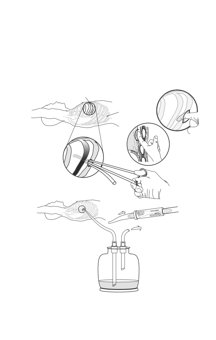
Procedure (Fig. 16-2).
ο In cases of tension pneumothorax, immediate decom-
pression with a large bore needle is lifesaving. An IV
catheter (14/16/18 gauge at least 2-3 inches in length) is
inserted in the midclavicular line in the second interspace
(approximately 2 fingerbreadths below the clavicle on the
adult male). Entry is confirmed by the sound of air passing
through the catheter. This must be rapidly followed by
the insertion of a chest tube.
ο In a contaminated environment, a single gram of IV
cefazolin (Ancef) is recommended.
ο If time allows, prep the anterior and lateral chest on the
affected side with povidone-iodine.
16.4
Thoracic Injuries
ο Identify the incision site along the anterior axillary line,
intersecting the 5th or 6th rib.
ο Inject a local anesthetic in the awake patient, if conditions
allow.
ο Make a transverse incision, 3-4 cm in length, along and
centered over the rib, carrying it down to the bone.
a
6th Rib Site
b
4
5
4
5
6
7
3
8
6
2
9
1
10
Anterior
Axillary Line
5
c
d
Lungs
4
5
6
6
7
e
4
5
6
7
2
3
8
Heimlich valve
1
9
10
OR
Suction/ Open
Chest tube
Fig. 16-2. Procedure for tube thoracostomy.
16.5
Emergency War Surgery
ο
Insert a curved clamp in the incision, directed over the
top of the rib, and push into the chest through the pleura.
A distinct pop is encountered when entering the chest and
a moderate amount of force is necessary to achieve this
entry. A rush of air out of the chest will confirm a tension
pneumothorax. Insertion depth of the tip of the clamp
should be limited by the surgeon’s hand to only 3 or 4 cm
to make sure that the clamp does not travel deeper into
the chest, resulting in damage to underlying structures.
ο
Spread the clamp gently and remove. The operator’s finger
is then inserted to confirm entry.
ο
Insert a chest tube (24 to 36 French) into the hole. All chest
tube side-holes must be in the chest. If no chest tubes are
available, an adult endotracheal tube may be used.
ο
Attach a chest tube to a Heimlich valve, sealed pleurovac,
or bottles. In a resource constrained environment, a cut-
off glove or Penrose drain may be attached to the end of
the chest tube.
ο
Secure the tube with suture, if possible, and dress to
prevent contamination.
Resuscitative Thoracotomy
ï Only indicated in penetrating chest injury in extremis
or with recent loss of vital signs.
ï These patients are generally unsalvageable, even with
unlimited resources and no other significant casualties.
ï If performed, a rapid assessment of injuries should be
made, and in the case of unsalvageable injuries, the
procedure should be immediately terminated.
Procedure
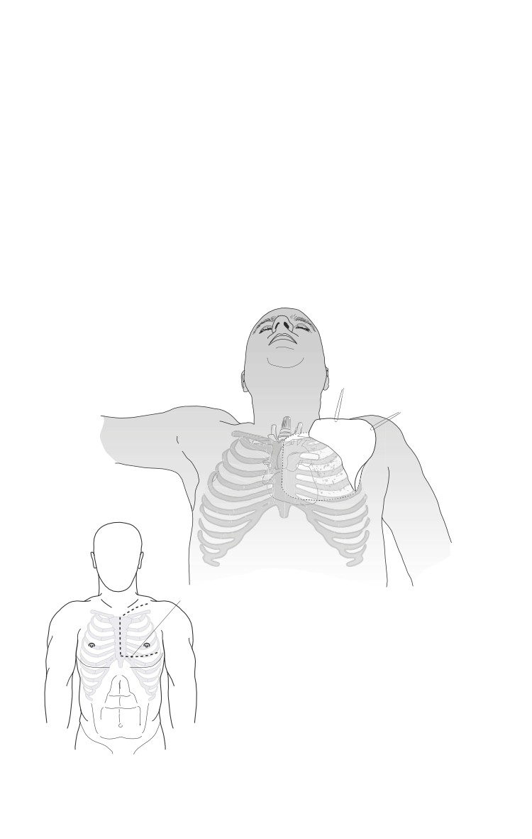
With the patient supine, make an incision in the left
inframammary fold starting at the lateral border of the
sternum extending to the midaxillary line (Fig. 16-3).
The procedure should be abandoned upon the discovery of
devastating injuries to the heart and great vessels.
16.6

Thoracic Injuries
If no injury is found in left
chest, rapidly extend the
incision across the midline,
crossing through the sternum
with a Lebsche sternum knife,
performing a mirror-image
thoracotomy (clamshell,
Fig. 16-4). When doing this
procedure you will cut across
both internal mammary
arteries, which will be a sig-
Fig. 16-3. Incision for resuscitative
nificant source of bleeding.
thoracotomy.
Elevating the anterior chest
wall will expose virtually all mediastinal structures.
Open the pericardium and assess the heart.
Priorities are to stop bleeding and restore central perfusion.
ο Holes in the heart and/or great vessels should be
temporarily occluded.
♦ Temporary occlusion can be achieved with fingers, side-
biting clamps or Foley catheters with 30 cc balloons.
Any other sterile device of opportunity is acceptable.
ο Major pulmonary hilar injuries should be cross-clamped
en masse.
ο Descending aorta located, cross-clamped, and cardiac
function restored via defibrillation or massage. (Make sure
to open the mediastinal pleura over the aorta to securely
apply the vascular clamp.)
ο If unable to restore cardiac function rapidly, abandon the
operation.
With successful restoration of cardiac function, injuries should
be more definitively repaired.
Subxiphoid Pericardial Window
Subxiphoid pericardial window should not be attempted
in an unstable patient. Unstable patients with penetrating
injuries suspicious for cardiac injury should undergo
immediate median sternotomy/thoracotomy.
16.7
lavicular Extension
udinal incision
h the skin and
exposing the
Thoracic Injuries
Place two stay sutures into the membrane and sharply incise
between them, with care to avoid the heart, opening the
pericardial sac and exposing the underlying beating heart.
Median Sternotomy
Indications.
ο Suspected cardiac injury in an unstable patient.
ο Positive pericardiocentesis/subxiphoid pericardial
window.
ο Suspected injury to the great vessels in the chest.
ο Suspected distal tracheal injury.
Procedure.
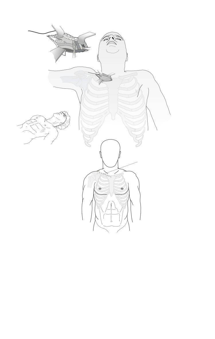
ο In the supine position, make a midline skin incision from
the sternal notch to just below the xiphoid.
ο Through blunt/sharp dissection, develop a plane for
several centimeters both superiorly and inferiorly beneath
the sternum.
ο Divide the sternum with a sternal saw or Lebsche knife.
Keep the foot of the knife/saw tilted up toward the
undersurface of the sternum to avoid cardiac injury. Bone
wax can be used to decrease bleeding on the cut edges of
the sternum.
ο Separate the halves of the sternum using a chest retractor.
ο Carefully divide the pericardium superiorly, avoiding the
innominate vein, exposing the heart and base of the great
vessels.
In general, exposure to the heart and great vessels is best
achieved through a median sternotomy. For proximal left
subclavian artery injuries, additional exposure (trap
door) may be necessary.
ο Close with wire suture directly through the halves of the
sternum, approximately 2 cm from the edge, or around
the sternum through the costal interspaces using wire
sutures.
ο Place one or two mediastinal tubes for drainage, exiting
through a midline stab wound inferior to the mediastinal
skin incision.
16.9
Other Approaches
Supraclavicular.
ο Indication.
♦ Mid to distal subclavian artery injury.
ο Procedure.
♦ Make an incision 2 cm above and parallel to the clavicle,
beginning at the sternal notch and extending laterally 8 cm.
Trap door (Fig. 16-5).
ο Indication.
♦ Proximal left subclavian artery injury.
ο Procedure.
on
Fig. 16-5. Trap door procedure.
Thoracic Injuries
♦ Perform supraclavicular approach as above.
♦ Perform a partial median sternotomy to the fourth
intercostal space.
♦ At the fourth intercostal interspace, incise the skin
laterally in the submammary fold to the anterior axillary
line.
♦ Divide the sternum laterally and continue in the 4th
intercostal space (ICS) to the anterior axillary line. The
internal mammary artery will be divided and must be
controlled.
♦ It may be necessary to either fracture or remove a section
of clavicle to gain adequate exposure of the proximal
left subclavian artery.
♦ Approach distal left subclavian artery injuries through
a supraclavicular incision.
Thoracoabdominal.
ο Indication.
♦ Combined thoracic and abdominal injuries.
ο Procedure.
♦ The resuscitative thoracotomy can be continued
medially and inferiorly across the costal margin into
the abdominal midline to complete a thoracoabdominal
incision.
♦ Alternatively, a separate abdominal incision can be
made.
♦ With right-sided lower chest injuries, the liver and
retrohepatic vena cava can be exposed well using a right
thoracoabdominal approach.
Specific Injuries
Vascular.
ο Initially, holes in vessels should be digitally occluded.
Stopgap measures include placing Fogarty or Foley
catheters, side-biting clamps, or in the case of venous
injuries, sponge sticks.
ο Total occlusion or clamping may temporarily be necessary
to allow resuscitation to continue and restore cardiac
function.
16.11
Emergency War Surgery
ο If cardiac function cannot be restored within 5 to 10
minutes, the procedure should be abandoned (on-the-table
triage).
ο Repair of vessels should follow the principles detailed in
vascular repair: attempting primary repair if possible, with
the use of prosthetics if primary repair is not feasible.
Consider shunting as an alternative.
Heart.
The usual result of high-velocity injuries to the heart is
irreparable destruction of the muscle.
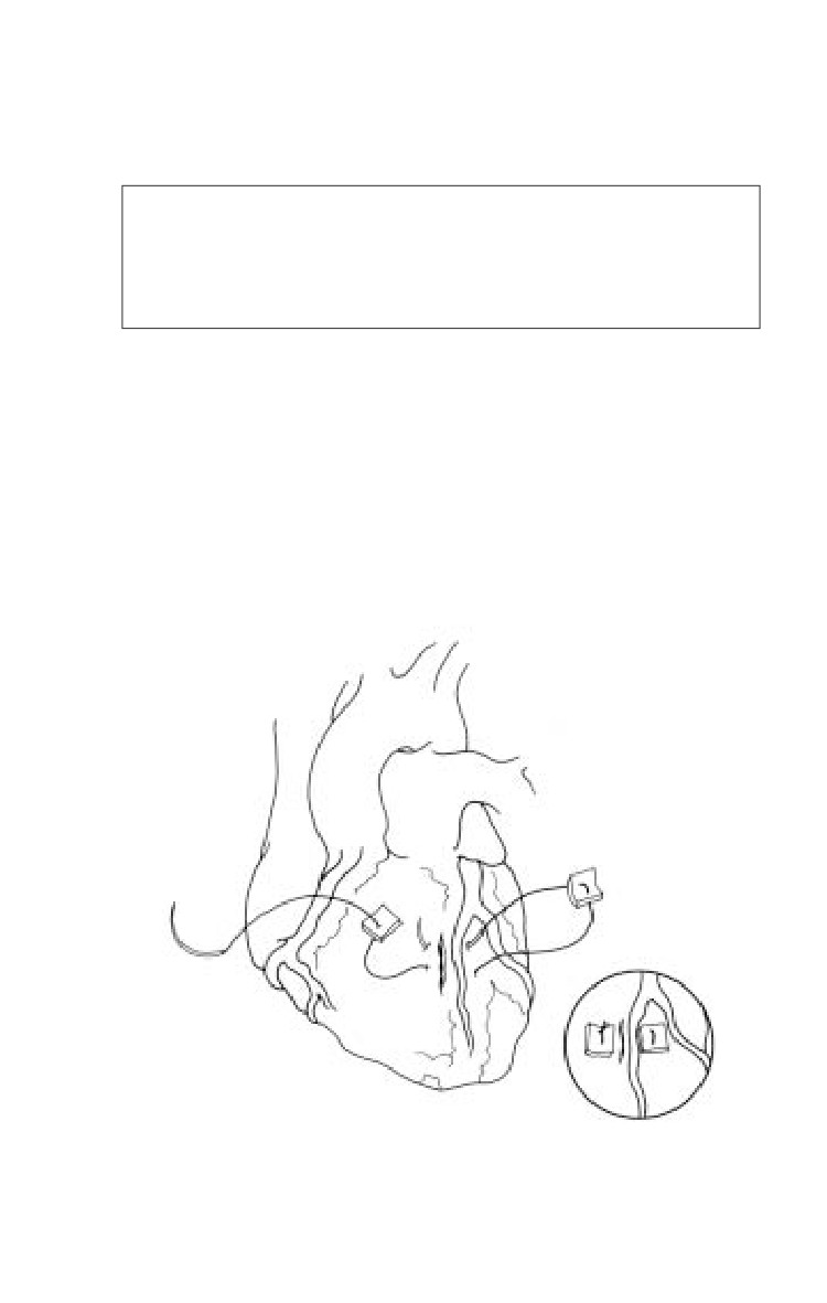
ο Isolated punctures of the heart should be exposed (opening
the pericardium) and occluded by finger pressure. Other
methods include the use of a Foley catheter or skin staples.
ο Use pledgeted horizontal mattress sutures (2-0 prolene)
on a tapered needle for definitive repair. Care must be
taken to avoid additional injury to coronary vessels.
Extreme care must be taken to avoid tearing the cardiac
muscle.
ο Atrial repairs may include simple ligature, stapled repair,
or running closures (Fig. 16-6).
ο Temporary inflow occlusion may prove helpful in repair.
ο More complex repairs are impractical without cardiac
bypass.
Lung.
ο Tube thoracostomy alone is adequate treatment for most
simple lung parenchymal injuries.
ο Large air leaks not responding to chest tubes or that do
not allow adequate ventilation will require open repair (see
tracheobronchial tree below).
ο Posterolateral thoracotomy is preferred for isolated lung
injuries. Anterior thoracotomy may also be used.
ο Control simple bleeding with absorbable suture on a
tapered needle. Alternatively, staples (TA-90) may be used
for bleeding lung tears.
16.12
Thoracic Injuries
ο Tractotomy: Open any bleeding tracts (through and
through lung penetrations) with a GIA stapler and ligate
bleeding points.
Do not simply close the entrance and exit points of
penetrating tracts in the lung. With positive pressure
ventilation, the risk is air embolism. The more central
the injury, the higher the risk.
ο Resection for bleeding may be indicated with severe
parenchymal injury. Anatomic resections are not indicated
and simple stapled wedge excisions recommended.
ο Uncontrolled parenchymal/hilar bleeding, or complex
hilar injuries with massive air leak should be controlled with
hilar clamping and repair attempted. Pneumonectomy is
performed as a last resort (90% mortality).
Fig. 16-6. Repair of penetrating cardiac injury.
16.13
Emergency War Surgery
Tracheobronchial tree.
ο Suspect the diagnosis with massive air leak, frothy
hemoptysis, and pneumomediastinum.
ο Confirm by bronchoscopy.
ο Airway control is paramount.
ο Median sternotomy is best approach.
ο Repair over endotracheal tube with absorbable suture --
may require segmental resection. Bolster with pleural or
intercostal muscle flap.
ο Temporizing measures include:
♦ Single lung ventilation.
♦ Control the airway through the defect.
Esophagus.
ο Isolated thoracic esophageal injuries are exceedingly rare.
They will usually be diagnosed incidentally associated
with other intrathoracic injuries.
ο Diagnostic clues include pain, fever, leucocytosis, cervical
emphysema, Hamman’s sign, chest X-ray (CXR) evidence
of pneumothorax, mediastinal air, and pleural effusion.
Contrast swallow may confirm the diagnosis.
ο Start IV antibiotics as soon as the diagnosis is suspected,
and continue post-op until fever and leucocytosis resolve.
This is an adjunctive measure only. Surgery is the
definitive treatment.
ο For stable patients in a forward location, chest tube
drainage and a nasogastric tube placed above the level of
injury is a temporizing measure. Ideally, primary repair is
performed within 6-12 hours of injury. Beyond 12 hours,
isolation of the injured segment may be necessary.
The preferred approach for intrathoracic esophageal
injuries is posterolateral thoracotomy; right for upper
esophagus and left for lower esophagus.
ο Locate the injury by mobilizing the esophagus. Primarily
repair with a single layer of 3-0 absorbable suture and cover
with pleural or intercostal muscle flap.
16.14
Thoracic Injuries
ο
Drainage with chest tubes (one apical, one posterior) is
recommended.
ο
If unable to primarily repair (as with a large segmental
loss or severely contaminated/old injury), staple above
and below the injury, place a nasogastric (NG) tube into
the upper pouch and place a gastrostomy tube into the
stomach. Drain the chest as indicated above. Complex
exclusion procedures are not indicated in a forward
operative setting.
ο
An alternative when the esophageal injury is too old for
primary repair is to close the injury over a large T-tube,
which converts the injury to a controlled fistula. The
mediastinum is then widely drained using chest tubes or
closed suction catheters placed nearby. After a mature
fistula tract is established, slowly advance the T-tube and
later the mediastinal drains can be slowly advanced.
Diaphragm.
ο All injuries of the diaphragm should be closed.
♦ Simple small lacerations (< 2 cm) should be reapprox-
imated with interrupted nonabsorbable 0 or 1-0
horizontal mattress sutures.
♦ Lacerations larger than 2 cm should be approximated
as above, then reinforced with a running suture to assure
an airtight closure.
♦ Care should be exercised in the central tendon area to
avoid inadvertent cardiac injury during the repair.
ο If there is significant contamination of the pleural space
by associated enteral injuries, anterior thoracotomy and
plueral irrigation and drainage with two well-placed chest
tubes should strongly be considered.
♦ Inadequate irrigation and drainage leads to a high
incidence of empyema, especially of the fungal variety.
16.15
Chapter 17
Abdominal Injuries
Introduction
Changing patterns of warfare together with improvements in
protective body armor combine synergistically to minimize
truncal trauma incidence, severity, and mortality, despite
increasingly lethal weapons systems. Despite these advances,
penetrating abdominal trauma still occurs and treatment of these
injuries will always be an important component of war surgery.
Trauma to the abdomen, both blunt and penetrating, can lead
to occult injury that can be devastating or fatal if not treated. In
the unstable patient with abdominal injury, the decision to
operate is usually straight forward and should be acted on as
soon as it is made. In a few rapidly hemorrhaging patients with
thoracoabdominal injuries, a rapid decision must be made as to
which cavity to enter first. This chapter addresses some of these
issues.
Penetrating injuries below the nipples, above the
symphysis pubis, and between the posterior axillary lines
must be treated as injuries to the abdomen and mandate
exploratory laparotomy.
Posterior truncal penetrating injuries from the tip of the
scapula to the sacrum may also have caused retroperitoneal
and intra-abdominal injuries. A low threshold for exploratory
laparotomy in these patients is warranted when there are not
other diagnostic modalities available.
Diagnosis of Abdominal Injury
Document a focused history to include time of injury,
mechanism of injury, previous treatments employed, and any
drugs administered.
17.1
Emergency War Surgery
Inspection of the chest and abdomen will be the most reliable
part of the physical examination, especially regarding
penetrating injuries.
Determine if the patient requires laparotomy, not the specific
diagnosis.
Indications for Laparotomy - Who, When, and Where
First imperative is to determine who needs surgery.
Patients who have
ο Penetrating abdominal wounds as described in box above.
ο Other penetrating truncal injuries with potential for
peritoneal penetration and clinical signs/symptoms of
intraperitoneal injury.
ο Blunt abdominal injuries presenting in shock.
When and Where.
ο When aeromedical evacuation is uncertain and will involve
substantial distance, unstable patients with life or limb
threatening circumstances should undergo laparotomy at
the nearest forward surgical team (FST).
ο Stable patients who can tolerate transport and delay of 6
hours or so, should undergo initial controlled resuscitation,
presurgical care (including antibiotics), and be transported
to the next level of care for surgery.
When the tactical situation is static, aeromedical evacuation
effective, and the distance between FST and combat support
hospital (CSH) or higher level hospitals is short, all casualties,
including those who are unstable, should bypass the FST and
be taken directly to a higher level hospital.
Diagnostic Adjuncts
Minimally invasive adjuncts to diagnosis—computed
tomography (CT) scan, diagnostic peritoneal lavage (DPL), and
ultrasound (US)—have been used to decrease the number of
negative laparotomies in stable, blunt abdominal trauma
patients in peace-time settings with good follow-up of patients.
Some have been used in lieu of laparotomy to evaluate those
with penetrating injuries, when the suspicion is high that no
intra-abdominal injury has occurred. This practice has the
17.2
Abdominal Injuries
potential of missing injuries. These diagnostic screening
procedures are primarily used in stable patients with a
mechanism of injury suggesting abdominal injury, but without
an obvious operative indication. They should be relied on only
when good follow up is possible. US and, to a limited extent,
DPL have some use in the unstable patient to indicate which
cavity should be entered first. US and DPL may also serve as
triage tools in the mass casualty situation.
Abdominal Ultrasound
Advantages: Noninvasive, may repeat frequently, quick, easy,
identifies fluid in the abdomen reliably.
Disadvantages: Operator dependant, may miss small
amounts of fluid associated with hollow-viscus injuries.
Sonography (focused abdominal sonography for trauma
[FAST]) has become an extension of the physical examination
of the abdomen and should be performed whenever available
and when abdominal injury is suspected.
ο 3.5 to 5 MHz curved probe is optimal.
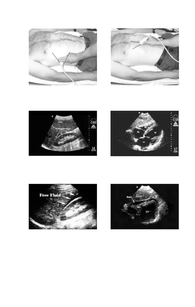
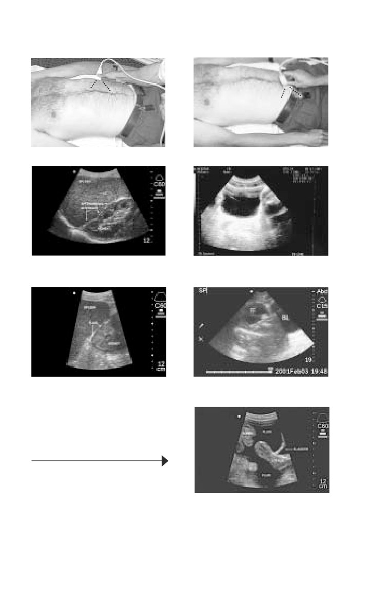
ο The abdomen is examined through four standard
sonographic windows.
A FAST examination assists the surgeon to determine the need
for laparotomy in blunt-injured patients but does NOT
identify specific injuries.
ο A FAST examination does not identify or stage solid organ
or hollow-viscous injury, but reliably identifies free
intraperitoneal fluid.
FAST aids in prioritization of penetrating injury patients for
the OR.
FAST aids in identifying which cavity to open first in patients
with thoracoabdominal injuries.
A FAST examination identifies pericardial fluid, and may
assist in the diagnosis of hemopneumothorax.
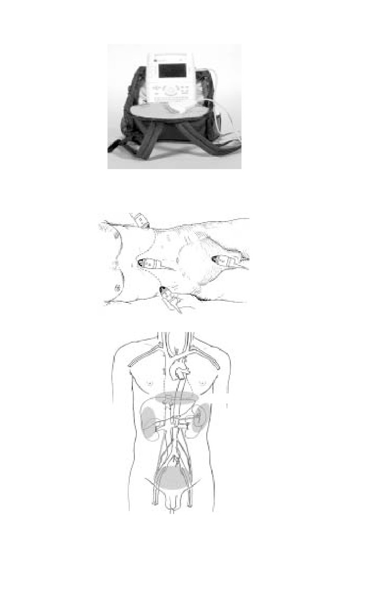
Ultrasound Views
A typical portable sonography device is shown in Fig. 17-1. The
standard locations for “sonographic windows” are shown in
Fig. 17-2. Examples of positive and negative sonographic
examinations are shown in Figs. 17-3 through 17-6.
17.3
Emergency War Surgery
Fig. 17-1. Typical sonography device.
a
b
Cardiac
LUQ
RUQ
Pelvic
Fig. 17-2 a,b. The standard four locations for sonographic windows.
17.4
Abdominal Injuries
a
a
b
b
normal
normal
c
c
abnormal
abnormal
Fig. 17-3 a,b,c. Normal and
Fig. 17-4 a,b,c. Normal and
abnormal negative sonographic
abnormal negative sonographic
examinations for the right upper
examinations for the cardiac
quadrant.
window.
17.5
Emergency War Surgery
a
a
b
b
normal
normal
c
c
abnormal
abnormal, male
d
Fig. 17-5 a,b,c. Normal and
abnormal negative sonographic
examinations for the left upper
quadrant.
Fig. 17-6 a,b,c,d. Normal and
abnormal negative sonographic
examinations for the pelvic
abnormal, female
window.
17.6
Abdominal Injuries
Diagnostic Peritoneal Lavage
DPL has been a mainstay of blunt abdominal trauma diagnosis
for many years. Unfortunately, in-theatre combat medical units
from Level 1 to Level 3 are not routinely outfitted with
microscopic laboratory functions to provide cell counts or
fluid enzyme determinations. Thus, the only reliable
information obtained from DPL is the aspiration of 10 cc of gross
blood. Gross blood aspiration is the most infrequently
positive criterion of DPL, and its value is probably supplanted
by FAST.
May be useful when US or CT are not available, or as triage
tool.
Requires laboratory for most sensitivity.
ο Blunt: Aspiration of 10 cc of gross blood, RBCs > 1,000,000/
mL, WBCs > 500/mL, fecal material.
ο Penetrating: not recommended to ruling out (R/O) injury
in penetrating combat wound.
May help determine which body cavity to enter first in an
unstable patient with truncal injury.
Advantages: Sensitive to small amounts of fluid, including
hollow-visceral leaks; fairly quick.
Disadvantages: Invasive, not repeatable, slower than US.
Kits allow Seldinger technique.
ο Arrow (AK-09000).
ο Baxter Lazarus-Nelson (MLNK9001).
Field Expedient substitution: Open technique with small,
vertical infraumbilical incision and any tubing (IV, straight
or balloon catheter). Cut at least a dozen extra side holes.
CT Scan
Advantages: Defines injured anatomy in stable patients.
Disadvantages: Slow; requires contrast use and equipment
availability; may miss small hollow organ leaks; requires
transport away from emergency care area; operator/
interpreter dependant; difficult to repeat.
17.7
Emergency War Surgery
Wound Exploration
Blast injuries and improvised explosive devices (IEDs) create
many low-velocity fragments that may penetrate the skin but
not the abdominal cavity. Operative wound exploration in
the stable patient with a normal or equivocal examination
can help determine the need for formal exploratory
laparotomy.
ο When possible wound exploration should be performed
in the operating room with adequate instruments and
lighting.
ο Finding the fragment in the abdominal wall precludes
laparotomy.
ο If the tract is not adequately identified or the fragment
seen on plain film cannot be identified, formal laparotomy
should be performed.
Operative Planning and Exposure Techniques
Give broad spectrum antibiotic pre-op, continue for 24 hours.
ο Redose short half-life antibiotics intraoperatively and
consider redosing antibiotics with large amounts of blood
loss.
Perform laparotomy through a midline incision.
ο When wide exposure is needed, extend the incision
superiorly just lateral to the xiphoid process and inferior
to the symphysis pubis.
Quickly pack all 4 quadrants while looking for obvious
injuries.
Control hemorrhage.
Assess physiologic status.
ο Considering casualty physiology, create operative plan to
control contamination and complete operation.
♦ Consider damage control (see Chapter 12, Damage
Control Surgery) early and often.
♦ If stabilized/improving, proceed with definitive surgery.
Identify all organ and hollow-viscus injuries.
17.8
Abdominal Injuries
Eviscerate the small bowel to increase workspace.
Divide the ligamentous attachments of the liver to improve
exposure in the right upper quadrant or upper midline.
Fold the left lateral segment of the liver down and to the right
to improve exposure at the gastroesophageal junction.
Improve exposure to the liver by extending the incision into
the inferior sternum and across into the lower right chest
(thoracoabdominal).
Stomach Injuries
The stomach is a vascular organ and will do well after almost
any repair.
ο Always enter the lesser sac to determine posterior wall injuries.
Encircle the distal esophagus with a Penrose drain to provide
traction and improve visibility in high midline injuries.
Minimally debride and primarily close stomach defects.
Duodenum Injuries
Injuries to the Duodenum are associated with massive upper
abdominal trauma. Early consideration for damage control
surgery should be considered (see Chapter 12, Damage Control
Surgery).
Missed injuries of the duodenum have devastating morbidity.
Bile staining or hematoma in the periduodenal tissues mandates
full exploration of the duodenum (Kocher maneuver).
Minor injuries can be repaired primarily.
Major injuries should be repaired if the lumen will not be
narrowed by more than 50%. Options for closing injuries of
greater than 50%:
ο Close duodenal wall around a tube duodenostomy.
♦ Use a No. 2-0 absorbable suture (Vicryl).
♦ Use the largest malecot catheter available.
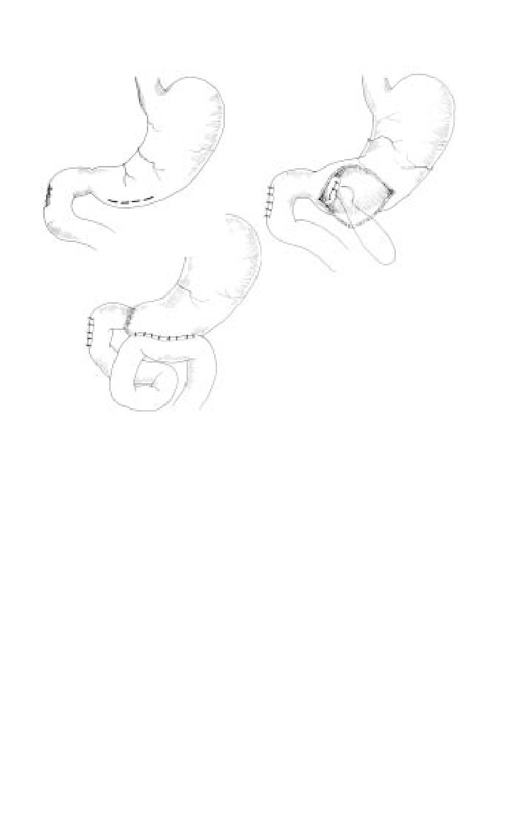
ο Bring up a Roux-en-Y jejunal limb and create an
anastamosis between the limb and the injury (Fig. 17-7).
ο The procedure of last resort is pancreaticoduodenectomy.
17.9
Emergency War Surgery
a
b
c
Fig. 17-7. (a) Ligation of pylorius, (b) Duodenal injury, (c) Roux-en-Y
anastomosis.
For major injuries, divert the gastric stream with a
gastrostomy and close the pylorus—two options:
ο Through a gastrotomy, ligate the pylorus with No. 0
absorbable suture.
ο Using a noncutting stapling device, staple but do not
divide the pylorus. Place a feeding jejunostomy for
nutrition at this controlled reconstruction.
Widely drain all injuries with closed-suction drains.
Any method used to close the pylorus will last only 14-21
days. The possibility of injury to the biliary and pancreatic
ducts should be considered when injuries involve the 2nd
portion of the duodenum or the pancreatic head.
Pancreas Injuries
Any injury to the pancreas/duct requires drainage.
17.10