Emergency War Surgery (2004) - page 11
Environmental Injuries
arising in the morning, attributed to increased
hypoxemia caused by altitude-induced sleep apnea.
♦ Anorexia.
♦ Nausea.
♦ Fatigue (weakness).
♦ General malaise.
♦ Decreased coordination.
♦ Dizziness or light-headedness.
♦ Oliguria.
♦ Emesis (vomiting).
♦ Lassitude.
♦ Insomnia: Sleep disturbances with periodic breathing
with recurrent apneic periods during sleep are usually
present, but are not necessarily a component of AMS.
ο
Diagnosis.
♦ Occurrence of a headache and at least one other sign/
symptom in an individual who ascended from low
(1,524 m or < 5,000 ft) to high altitude, or high altitude
to higher altitude in the previous 24-48 hours.
♦ Differential diagnosis includes viral gastroenteritis,
hangover, exhaustion, dehydration, carbon monoxide
poisoning, and HACE.
♦ Presence of neurologic symptoms such as incoord-
ination, ataxia, and excessive lethargy or cognitive
dysfunction is indicative of progression to HACE, which
requires immediate therapeutic intervention.
ο
Prophylaxis for AMS.
♦ Gradual acclimation.
◊ Staged ascent: Soldiers ascend to intermediate
altitudes and remain there for 3 or more days before
ascending further.
◊ Graded ascent: Limits daily altitude gain to allow
partial acclimation. Sleep altitude is most important.
Have soldiers spend 2 nights at 2,743 m (9,000 ft) and
limit the sleeping altitude to no more than 305 m
(1,000 ft) per day above previous night’s sleep
altitude.
◊ Combination of both staged and graded ascent is the
safest and most effective prevention method.
29.25
Emergency War Surgery
♦ Diet: High carbohydrate diet (< 70% of total energy
intake as carbohydrates) (stimulation of ventilation
through increased carbon dioxide produced from
metabolism of carbohydrates).
♦ Acetazolamide, 250 mg qid or 500 mg bid po, starting
48 hours before ascent, continuing for 48 hours after
ascent. Side effects include peripheral paresthesias,
fatigue, increased urination (polyuria), and altered taste
imparted to carbonated beverages. It prevents AMS in
50%-75% of soldiers and reduces symptoms in most
others. Short-term use when changing altitude
significantly (400 m). Contraindicated in sulfa allergy.
♦ Dexamethasone, 4 mg qid po is the prophylaxis of choice
in sulfa-allergic individuals. Dexamethasone does not
aid acclimatization and effects are gone when it is
stopped. Dexamethasone +/- acetazolamide is also
prophylaxis of choice for missions of a rapid, high (over
4,000 m [13,000 ft]), short-duration profile (raids,
rescues).
♦ Cyanosis: Oxygen 2-6 L/min. Do not delay descent.
ο
Treatment.
♦ AMS alone does NOT mandate descent.
♦ Remain at the same elevation; do not ascend until
symptoms abate.
♦ Acetazolamide, 125 mg qid to 500 mg, tid, po—do not
use in patients with sulfa allergies. (If already receiving
a preventive dose of acetazolamide (1,000 mg/d) and
still symptomatic, 500 mg can be added with caution.
♦ Dexamethasone in doses of 2-4 mg q6h (has the same
potentially serious side effects as when used as a
prophylaxis). Symptoms may recur when medication
stopped.
♦ Oxygen by nasal cannula 2-6 L/min (severe headache).
♦ Do NOT advance sleeping altitude.
♦ Symptomatic treatment with ASA, acetaminophen,
prochlorperazine for nausea and vomiting 5-10 mg tid-
qid, po or IM, or 25 mg bid prn also stimulates
respiration; ibuprofen for headache.
♦ Minimize utilization of sleeping agents at altitude; they
29.26
Environmental Injuries
can worsen illness. Acetazolamide for sleep disorders,
250 mg qid or tid po. Temazepam for insomnia 30 mg
qhs po; triazolam for insomnia 0.125-0.25 mg qhs po.
Short-term use only. Possible short-term memory loss.
High-altitude pharyngitis and bronchitis.
ο Common condition occurring after 2-3 weeks at altitude.
ο Common at altitudes over 5,486 m (18,000 ft).
ο Sore throat, chronic cough, and severe cough spasms
(severe enough to cause rib fractures).
ο Environmental, from breathing cold dry air.
ο Altitude-induced tachypnea aggravates the problem.
ο Cold-induced vasomotor rhinitis, especially at night,
stimulates mouth breathing and also aggravates problem.
ο Usually not caused by infection, although infection can
occur.
ο Patient will not have dyspnea at rest.
ο Symptomatic treatment with lozenges, mild cough
suppressant, and decongestant nasal sprays. Personnel can
use a mask or a porous, breathable silk balaclava as a
mouth covering to reduce respiratory heat and moisture
loss.
ο Maintain hydration.
High-altitude peripheral edema.
ο Altitude-related edema of hands and face.
ο Hypoxia-induced retention of sodium and water.
ο Not considered related to AMS/HACE edema-spectrum
or HAPE.
ο Decreased urine output and weight gain of 2.7-5.4 kg
(6-12 lb) over several days; most evident upon awakening.
ο Diagnosis based on association of characteristic peripheral
edema with ascent to high altitude; recurs consistently with
repeat ascents; more common in females.
ο Differential diagnosis includes cardiogenic edema, allergic
reactions, and edema of the upper extremities caused by
pack straps or binding by tight clothes.
ο Prophylaxis includes salt restriction. The acetazolamide
regimen used to prevent AMS is often successful in
preventing peripheral edema.
29.27
Emergency War Surgery
ο Treatment with diuretics (one 20-40 mg dose of furosemide,
or 250 mg of acetazolamide every 8 h for 3 doses) and salt
restriction.
High-altitude retinal hemorrhage (HARH).
ο Bleeding from retinal vessels during altitude exposure.
One of the manifestations of hypoxia-induced retinopathy.
ο Caused by BP “surges” within the distended vessels.
ο Usually asymptomatic; normally does not adversely affect
military operations; however, can affect an individual
soldier’s vision.
ο Hemorrhages are self-limiting and resolve in 1-2 weeks
after descent.
Thromboembolic events.
ο
Increased possibility of thromboembolic event with ascent
to high altitude: thrombophlebitis, deep venous thrombosis,
pulmonary embolus, transient ischemic attacks (TIAs), and
stroke.
ο
Probably result from hypoxia-induced polycythemia and
clotting abnormalities but also may result from environ-
mental and mission factors such as dehydration, cold, and
venous stasis caused by prolonged periods of inactivity
during inclement weather or by constriction of tight-fitting
clothing and equipment.
ο
Unusual below 4,267 m (14,000 ft). At very high and
extreme altitudes (> 4,200 m [13,700 ft]) these events are
not uncommon, and thrombophlebitis appears to be
relatively common.
ο
Clinical manifestations are similar to manifestations of
thromboembolic events at low altitude, except for their
occurrence in young and otherwise healthy personnel.
ο
Prevention relies on reducing the risk factors by main-
taining adequate hydration and warmth and by avoiding
conditions that might cause venous stasis.
ο
Evacuation to lower altitude is required. Treatment follows
standard treatment guidelines, including appropriate anticoa-
gulation. In the field setting, fractionated heparin (one dose
of 250 IU/d) can be used prior to and during evacuation.
Subacute mountain sickness.
29.28
Environmental Injuries
ο Prolonged deployment (weeks to months) to elevations
above 3,658 m (12,000 ft).
ο Common manifestations include sleep disturbances,
anorexia, weight loss, fatigue, daytime somnolence, and
subnormal mentation.
ο Caused by failure to acclimatize adequately.
ο Some relief of symptoms obtained from low-flow oxygen
and from acetazolamide.
ο Evacuate to lower altitude as soon as practical.
ο Some degree of immune suppression and poor wound
healing occurs in personnel at very high and extreme
altitudes. Injuries resulting from burns, ballistics, and
physical trauma should be considered more clinically
significant at high altitude.
High-altitude pulmonary edema.
ο
Potentially fatal, noncardiogenic pulmonary edema.
ο
Occurs in < 10% of personnel ascending above 3,700 m
(12,000 ft).
ο
Onset 2-4 days after rapid ascent to altitudes greater than
2,438 m (8,000 ft).
ο
Repeated ascents and descents above 3,700 m (12,000 ft)
increase susceptibility.
ο
Risk factors.
♦ Moderate to severe exertion.
♦ Cold exposure.
♦ Anxiety.
♦ Young age.
♦ Male sex.
♦ Obesity (possibly).
ο
Early symptoms (pulmonary edema).
♦ Nonproductive cough.
♦ Rales (few).
♦ Dyspnea on exertion.
♦ Fatigue.
♦ Weakness with decreased tolerance for physical activity
and increased time for recovery after physical exertion.
♦ Resting tachycardia and tachypnea greater than induced
by altitude alone.
29.29
Emergency War Surgery
♦ Once symptoms appear, HAPE can progress very
rapidly (< 12 hours) to coma and death.
♦ Nail beds and lips may be more cyanotic than other unit
members.
ο
Progressing pulmonary edema.
♦ Productive cough of frothy and sometimes pink or
bloodstained sputum.
♦ Rales more numerous and widespread.
♦ Wheezing may develop.
♦ Lung sounds become audible even without stethoscope,
especially when individual is supine.
♦ Orthopnea may occur (< 20%).
♦ Progressive hypoxemia causes dyspnea and cyanosis.
♦ Arterial blood gas (if available) documents hypoxemia,
hypocapnia, and slight increase in pH.
♦ Mental status deteriorates with progressive confusion
and sometimes vivid hallucinations.
♦ Obtundation, coma, and death occur without treatment.
♦ Subfebrile temperature < 38°C (100.5°F) and a mild
increase in white blood cell count may be present.
♦ Dyspnea at rest.
♦ Marked hypoxia by oximetry.
♦ Dyspnea at rest and cough should be considered to
be the onset of HAPE.
DELAY IN TREATMENT OF PROGRESSIVE PULMONARY EDEMA AT
ALTITUDE USUALLY RESULTS IN DEATH.
ο Treatment.
♦ Depends on severity.
♦ Immediate descent is mandatory! Descent of even a few
hundred meters (300-1,000 m) can be helpful or even
lifesaving in severe cases.
♦ Mortality can approach 50% if descent cannot be
accomplished rapidly.
♦ Oxygen by cannula 2-6 L/min (mild), or by mask 4-6
L/min (moderate and severe). DO NOT DELAY
DESCENT!
29.30
Environmental Injuries
♦ Portable fabric hyperbaric chamber may be lifesaving—
Gamow bag/Certec SA.
♦ Nifedipine, 10 mg tid sublingually, or 20 mg po. A
second 10-mg, sublingual dose can be administered in
15-20 minutes if no improvement in symptoms is
apparent, followed by 30 mg qid.
Nifedipine should not be used in lieu of descent,
supplemental oxygen, or treatment in a hyperbaric bag.
It may be used in conjunction with other therapies.
♦ Immediate descent to lower elevation; if symptoms
resolve, wait at least 72 hours before attempted return
to previous elevation.
Neither furosemide nor morphine sulfate should be used
in the treatment of HAPE unless other more effective
treatment options are not available.
♦ Treatment after descent, at an MTF, is directed toward
ensuring adequate oxygenation and reducing pulmonary
artery pressure; includes bed rest, supplemental oxygen,
and nifedipine.
♦ Invasive diagnostic procedures such as bronchoscopy
or pulmonary artery catheterization are NOT indicated
unless clinical course deteriorates and the diagnosis is
in doubt. Endotracheal intubation is seldom necessary.
ο HAPE Prophylaxis.
♦ Nifedipine, 20 mg tid, po, 24 hours before ascent,
continuing 72 hours after ascent.
High-altitude cerebral edema.
ο Onset following ascent is highly variable and occurs later
than either AMS or HAPE. Mean duration of onset 5 days
with a range of 1-13 days.
ο Incidence lower than AMS or HAPE (< 1% of individuals
making rapid ascent).
ο Potentially fatal, uncommon (< 2% above 3,700 m). Can
occur as low as 2,430 m (8,000 ft) but vast majority of cases
29.31
Emergency War Surgery
above 3,600 m (12,000 ft). Untreated HACE can progress
to death over 1-3 days or become more fulminant with
death occurring in < 12 hours.
ο
Exacerbation of unresolved, severe AMS.
ο
Most often occurs in people who have AMS symptoms
and continue to ascend.
ο
Signs and symptoms.
♦ Most signs and symptoms are a manifestation of
progressive cerebral edema.
♦ Early signs resemble AMS (these symptoms are not
invariably present).
♦ Severe headache
♦ Nausea
♦ Vomiting.
♦ Extreme lassitude.
ο
Progressing signs.
♦ Mental status changes: Confusion, disorientation,
drowsiness, and impaired mentation.
♦ Truncal ataxia (swaying of upper body, especially when
walking). As the edema progresses, soldier may also
exhibit an ataxic gait in addition to the truncal ataxia.
♦ Soldier appears withdrawn, and behavior is mistakenly
attributed to fatigue or anxiety.
♦ Cyanosis and general pallor are common.
♦ Symptoms of HAPE.
ο
Untreated HACE.
♦ Variety of focal and generalized neurologic abnormalities
may develop: visual changes, anesthesias, paresthesias,
clonus, pathological reflexes, hyperreflexia, bladder and
bowel dysfunction, hallucinations, and seizures.
♦ Papilledema may be present in up to 50% of the soldiers,
but is NOT universal.
ο
Coma.
Ataxia at altitude is HACE.
ο
Prophylaxis.
29.32
Environmental Injuries
No definitive evidence; however, due to similarity with
AMS, prophylactic measures for HACE include use of
staged or graded ascent, high carbohydrate diet, and use
of acetazolamide.
ο
Treatment.
♦ Immediate descent is mandatory. Definitive treatment
of HACE is immediate descent. In general, the greater
the descent the better the outcome. Descent of more than
300 m (1,000 ft) may be required for clinical improvement,
and descents to altitudes of less than 2,500 m (8,000 ft)
is optimal.
♦ If descent is delayed, treatment with a portable cloth
hyperbaric chamber may be lifesaving. May require at
least 6 hours of pressurization in chamber.
♦ Oxygen by mask or cannula 2-6 L/m; should not be
used as a substitute for descent.
♦ Dexamethasone, 4-8 mg initially and then 4 mg qid,
po, IV, or IM. DO NOT DELAY DESCENT! Few side
effects if used only 3-4 days.
♦ Loop diuretics and osmotic diuretic agents, such as
mannitol, urea, and glycerol, have been suggested, but
there is little experience with them in this role. Careful
attention is required before diuretics are used.
Individual may have altitude-induced decrease in
intravascular volume concomitant with cerebral edema.
♦ Hospital management consists of supplemental oxygen
(if needed to maintain arterial oxygen levels), supportive
care, and possibly diuretics. Comatose patients may
require intubation and bladder catheterization.
HACE and HAPE often coexist. Individuals with HACE
will often have HAPE; however, most individuals with
HAPE do not have concomitant HACE.
29.33
Chapter 30
Radiological Injuries
The reader is strongly advised to supplement material
in this chapter with the following two references:
1. Medical Management of Radiological Casualties
Handbook, 2003, Armed Forces Radiobiology Research
Institute, Bethesda, MD.
2. Medical Management of the Acute Radiation Syndrome:
Recommendations of the Strategic National Stockpile.
Radiation Working Group, Strategic National
Stockpile (Annals of Internal Medicine, 15 June 2004).
Introduction
Radiological casualties on the battlefield may occur with
improvised or conventional nuclear devices or radiological
dispersal devices (“dirty bombs”) (Table 30-1).
Conventional nuclear weapons.
ο The relative casualty-causing potential depends primarily
on four factors:
♦ Yield of the weapon.
♦ Height of burst.
♦ Environmental conditions in which the detonation
occurs.
♦ Distribution and shielding of troops in the target area.
ο A nuclear detonation generally causes injuries with the
following distribution:
♦ Blast injury: 50%.
♦ Thermal injury: 35%.
♦ Ionizing radiation injury.
◊ Initial: 5%.
◊ Residual: 10%.
30.1
Emergency War Surgery
A radiological dispersal device (RDD) is any device, including
any weapon or equipment, other than a nuclear explosive
device, specifically designed to spread radiation.
ο RDDs contaminate conventional casualties with radionuc-
lides, complicating medical evacuation.
ο RDDs are ideal weapons for terrorism and are used to
intimidate and deny access to an area by spreading
radioactive material.
Table 30-1. Radiological casualties.
Weapon Effect
Weapon Yield (Kiloton)/Distance (Meters)
1 kt
10 kt
100 kt
1,000 kt
Blast (50% casualties)
140 m
360 m
860 m
3,100 m
Thermal radiation
370 m
1,100 m
3,190 m
8,020 m
(50% deep burns)
Ionizing radiation
600 m
950 m
1,400 m
2,900 m
(50% immediate transient
ineffectiveness)
Ionizing radiation
800 m
1,100 m
1,600 m
3,200 m
(50% lethality)
Triage
Different from conventionally injured patients, because
survivable radiation injury is not manifested until days to
weeks after exposure.
ο Based primarily on conventional injuries, then modified
by radiation injury level.
ο Make a preliminary diagnosis of radiation injury only for
those with exposure symptoms, such as nausea, vomiting,
diarrhea, fever, ataxia, seizures, prostration, hypotension.
ο Radiation patient triage classifications.
♦ Delayed: casualties with only radiation injury, without
gross neurological symptoms (ataxia, seizures, impaired
cognition). For trauma combined with radiation injury,
all surgical procedures must be completed within 36-
48 hours of radiation exposure, or delayed until at least
2 months after the injury.
30.2
Radiological Injuries
♦ Immediate: those requiring immediate lifesaving
intervention. Pure radiation injury is not acutely life-
threatening unless the irradiation is massive. If a
massive dose has been received, the patient is classified
as Expectant.
♦ Minimal: buddy care is particularly useful here.
Casualties with radiological injury should have all
wounds and lacerations meticulously cleaned and then
closed.
♦ Expectant: receive appropriate supportive treatment
compatible with resources; large doses of analgesics as
needed.
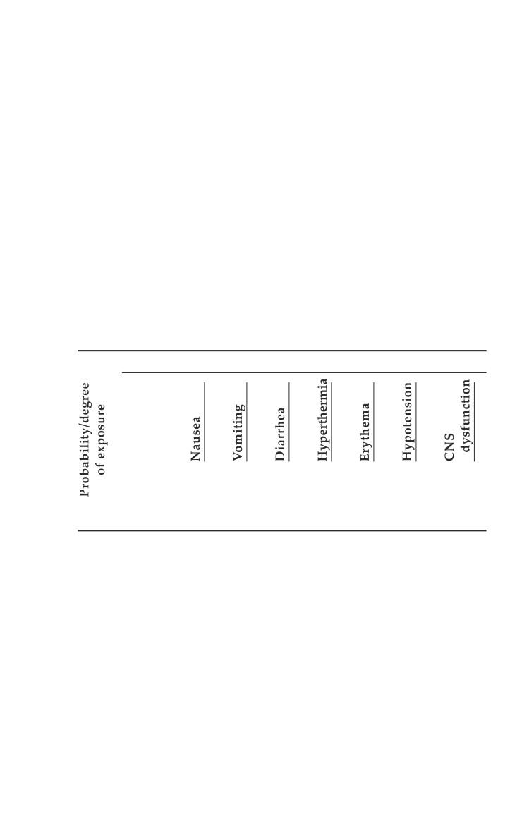
Table 30-2 provides medical aspects of radiation injuries.
Table 30-2. Medical aspects of radiation injuries.
Signs and Symptoms
Unlikely
-
-
-
-
-
-
-
Probable
++
+
+/-
+/-
-
-
-
Severe
+++
+++
+/+++ +/+++
-/++
+/++
-/++
The lethal dose of radiation, which will kill 50% of a population
within 60 days of exposure, is called LD50/60. The LD50/60 is
approximately 3-4 Gray (Gy) for a population with radiation
injury alone and with no significant medical care. The LD50/60
for a population with radiation injury alone and the best
available medical care (including antiemetics, antivirals,
antibiotics, hematopoietic cytokines, and transfusion) may be 6
Gy or more. Combined injuries with radiation and trauma and/
or burns will markedly lower the LD50.
30.3
Emergency War Surgery
Significant medical care may be required at 3-5 weeks for 10%-
50% of personnel. Anticipated problems should include
infection, bleeding, fever, vomiting, and diarrhea. Wounding
or burns will markedly increase morbidity and mortality.
Treatment.
ο Fluid and electrolytes for GI losses.
ο Cytokines for immunocompromised patients (follow
granulocyte counts).
ο Restricted duty. No further radiation exposure, elective
surgery, or wounding. May require delayed evacuation
from theater during nuclear war IAW command guidance.
ο If there are more than 1.7 • 109 lymphocytes per liter, 48
hours after exposure, it is unlikely that an individual has
received a fatal dose.
Patients with low (300-500) or decreasing lymphocyte counts,
or low granulocyte counts, should be considered for cytokine
therapy and biological dosimetry using metaphase analysis
where available.
Asymptomatic patients with lethal radiation dose may
perform usual duties until symptomatic.
Potential Injuries
Thermal/flash burns or thermal pulse burns are caused
directly by infrared radiation. Close to the fireball, the thermal
output is often so great that everything is incinerated, and
even at great distances, thermal/flash burns will occur (see
Chapter 28, Burns, for management).
ο Burn mortality rates associated with radiation exposure
are significantly higher due to bone marrow suppression
and infection (a 50% TBSA burn associated with radiation
exposure has a mortality of 90%).
Blast injuries associated with a nuclear detonation include:
ο Direct blast wave overpressure forces measured in terms
of atmosphere overpressure.
ο Indirect blast wind drag forces, measured in terms of wind
velocity, which may displace large objects such as vehicles
or cause the collapse of buildings.
30.4
Radiological Injuries
Radiation injuries are due to ionizing radiation released both
at the time of the nuclear detonation and for a considerable
time afterward. The two types of radiation released are
electromagnetic (gamma) radiation and particulate (alpha,
beta, and neutron) radiation.
ο Alpha particles can be shielded against by clothing.
ο Beta particles shielding requires solid materials, like a wall.
ο Gamma and neutron radiation are the most biologically
active, and require lead equivalent shielding for protection.
ο Fission products are the major radiation hazard in fallout,
because a large number emit penetrating gamma radiation.
This can result in injuries, even at great distances.
ο Fallout causes whole body irradiation from gamma-
emitting isotopes, because they do not actually have to be
on a person’s skin to cause damage.
Flash blindness may occur as the result of a sudden
peripheral visual observation of a brilliant flash of intense
light energy. Retinal burns may also occur and result in
scarring and permanent altered visual acuity.
Treatment of Combined Injuries
Following the detonation of a nuclear device, the majority of
resulting casualties will have sustained a combination of blast,
thermal, and radiological injuries.
The usual methods of treatment for blast injuries must be
modified in those casualties simultaneously exposed to
ionizing radiation.
Traditionally, combat wounds are left open. However, wounds
left open to heal by secondary intention in the irradiated
patient will serve as a nidus of infection. Wounds exposed to
ionizing radiation should be debrided and closed at a second-
look operation within 36-48 hours.
Hypotension should always be assumed to be hypovolemia
and not due to radiologic injury.
Hyperthermia is common.
Radiological injuries increase the morbidity and mortality of
injuries due to compromise of the normal hematopoietic and
30.5
Emergency War Surgery
immune responses to injury. Surgical procedures may need
to be delayed during bone marrow suppression if at all
possible.
Potassium iodide may be used for prevention of thyroid
uptake of radioisotopes after nuclear reactor accidents.
Chelating agents may be used to eliminate metals from the
bloodstream before they reach target organs.
Mobilizing agents are used to increase the excretion of internal
contaminants.
Prussian blue is used to remove radionuclides from the
capillary bed surrounding the intestine and prevents their
reabsorption. Delay until patient is stable. Treat ABCs first.
Decontamination
No healthcare provider has ever been injured with radiation
while performing ABCs on a radiation victim.
Removal of the casualty’s clothing can remove as much as
90% of the radiological contamination.
The first priority of surface decontamination should be to
open wounds, then other areas.
ο To prevent rapid incorporation of radioactive particles,
wounds should be copiously irrigated with normal saline
for several minutes.
ο The eyes, ears, nose, mouth, areas adjacent to uncontam-
inated wounds, hair, and remaining skin surface should
be decontaminated with soap and water.
ο Personnel providing decontamination must protect
themselves from ionizing radiation exposure with:
♦ Protective outer clothing.
♦ Aprons, gloves, and masks.
Amputation should be seriously considered when the
contamination burden is great and severe radionecrosis is likely.
Logistics of Casualty Management
If nuclear weapons are employed within the theater, the
entire medical evacuation and treatment system will be
severely overburdened and some system of classification and
sorting of casualties must be added to the normal procedures
of evacuation and hospitalization.
30.6
Radiological Injuries
Patients entering a medical treatment facility should be
routinely decontaminated if monitoring for radiation is not
available.
These two requirements, the sorting of casualties and the
holding of the excess numbers, must be planned for and
drilled as part of the normal organization and operation of
the health service support system in a theater of operations
where radiation exposure potential is high.
30.7
Chapter 31
Biological Warfare Agents
Introduction
Biological warfare (BW) agents infect the body via the same portals
of entry as infectious organisms that occur naturally. These include
inhalation into the respiratory tract, ingestion into the GI tract, and
absorption through mucous membranes, eyes, skin, or wounds. Most
BW agents will enter the body through inhalation. Usually, the
disease produced by a BW agent will mimic the naturally occurring
disease, but the clinical presentation can be different if delivery of
an agent occurs through a portal that differs from the natural portal.
Detection
Compressed epidemiology with record numbers of sick and
dying in a short time.
High attack rates (60%-90%).
High incidence of pulmonary involvement when usual form
of infection is not (eg, anthrax).
Incidence of a particular disease in an unlikely location.
Increased deaths of animals of all species.
Near simultaneous outbreaks of several different epidemics
at the same site.
Biological Identification Detection System or standoff BW
detectors alarming.
Direct evidence of an attack such as contaminated or
unexploded munitions.
Diagnosis
The first indication of an attack may be when large
numbers of patients present with the same constellation
of signs and symptoms, especially for a disease that is
not endemic to the area of operations.
31.1
Emergency War Surgery
Rapid diagnostic tests may be available in forward areas to
assist clinicians in early diagnosis:
Isolation of the etiologic agent can occur within 1-2 days for
some agents.
Enzyme-linked immunosorbent assays (ELISA).
Genome detection by polymerase chain reaction (PCR).
Antibody detection.
Prevention and Protection
Immunizations: Anthrax, and in specific scenarios, smallpox
and plague.
ο Pre- or postexposure chemoprophylaxis—anthrax, plague,
Q fever, and tularemia. Chemoprophylaxis for anthrax is
presently FDA-approved for postexposure only.
♦ Investigational new drugs exist for the treatment of
Argentine hemorrhagic fever, botulinum toxin, Q fever,
Rift Valley fever, Venezuelan Equine Encephalitis (VEE),
and tularemia.
Protective clothing and mask.
Decontamination —Personnel, Equipment, and Clothing
Mechanical decontamination removes, but not necessarily
neutralizes, the BW agent.
ο Brushing to ensure loosening of the BW agent from the
surface.
ο Filtration and chlorination of drinking water to remove
organisms.
Chemical decontamination renders BW agents harmless
through the use of disinfectants.
ο Soap and water followed with copious rinsing with water
is often sufficient.
ο For patients requiring urgent decontamination, biologic
agents are neutralized within 5 minutes when contam-
inated areas are washed with a 0.5 % hypochlorite solution
(1 part household bleach mixed with 9 parts water).
ο Do not use hypochlorite in the eyes, abdominal cavity,
or on nerve tissue.
ο A 5% hypochlorite solution (ie, household bleach) may be
used to decontaminate clothing or equipment.
31.2
Biological Warfare Agents
Physical decontamination such as heat and solar ultraviolet
(UV) radiation.
ο Dry heat for 2 hours at 160°C.
ο Autoclaving at 120°C under 1 atm of overpressure for 20
minutes.
ο UV radiation difficult to standardize.
Dry biological agents can be a hazard through secondary
aerosolization; but adequate liquid decontamination will
prevent this hazard. There is no vapor hazard, and special
protective masks are generally not required for surgical
personnel.
Infection Control
Infection control procedures should be reinforced for situations
involving BW agents. Standard precautions are appropriate for
BW agents once they have been identified. For an undifferen-
tiated febrile illness following a BW agent attack:
Place patients together in an isolated setting such as a
designated tent or other structure.
Surgical masks may be placed on patients when isolation is
not possible.
Employ respiratory droplet precautions along with standard
precautions until diseases transmissible by droplet (such as
plague and smallpox) have been excluded.
Medical Evacuation
If plague, smallpox, and the hemorrhagic fevers can be
excluded, patients may be evacuated using standard
precautions and the disease-specific precautions.
Plague and smallpox are internationally quarantinable
diseases (IQDs). Do not evacuate patient across
international borders unless authorized by the theater
surgeon.
Isolation precautions should be added to standard precautions.
Immediately upon diagnosing patients with smallpox, the
line and medical chain of command must be notified.
Observe strict quarantine.
31.3
Emergency War Surgery
ο
Standard and respiratory droplet isolation precautions.
♦ Standard precautions.
◊ Hand washing after patient contact.
◊ Use of gloves when touching blood, body fluids,
secretions, excretions, and contaminated items.
◊ Use of mask, eye protection, and gown during
procedures likely to generate sprays of blood, body
fluids, secretions, or excretions.
◊ Handle contaminated patient-care equipment and
linen in a manner that precludes transfer of micro-
organisms to individuals or equipment.
◊ Practice care when handling sharps and use pocket
mask or other ventilation device when ventilating
the patient.
◊ Place patient in private room when possible. Limit
the movement or transfer of patient.
♦ Droplet precautions.
◊ Standard precautions plus:
Place patient in private room or with someone
with the same infection. If not feasible, maintain
at least 1 m distance between patients.
Use of a mask when working within 1 m of patient.
Mask the patient if he/she needs to be moved.
ο
All contacts should be vaccinated within 7 days of
exposure and quarantined together for at least 17 days
following the most recent exposure.
Hemorrhagic fevers—Hanta, Ebola, Lassa, Rift Valley, HFRS
Except for yellow fever, quarantine is not mandatory;
however, person-to-person transmission is possible, therefore,
universal precautions are recommended.
Medical evacuation may result in increased morbidity and
mortality, thus treatment at local MTFs is preferred.
When necessary, patients may be evacuated using universal
and respiratory droplet isolation precautions.
31.4
Biological Warfare Agents
Biological Agents
There are two biological toxins that are potential BW agents:
botulinum and ricin (see Table 31-1).
Table 31-1
Biological Toxin Signs/Symptoms
Medical Management
Botulinum
Cranial nerve palsies
Antitoxin/supportive care
Paralysis
Respiratory failure
Ricin
Fever, cough, SOB
Arthralgias, pulmonary Nonspecific/Supportive care
edema
Bacterial Agents
The bacteria or rickettsia most often considered to be potential
BW threat agents include Bacillus anthracis (anthrax), Brucella
sp. (brucellosis), Vibrio cholerae (cholera) Burkholderia mallei
(glanders), Yersinia pestis (plague), Francisella tularensis
(tularemia), and Coxiella burnetii (Q Fever) (see Table 31-2).
Table 31-2
Bacterial Signs/Symptoms
Medical Management
Anthrax
Fever, malaise, cough, SOB,
Ciprofloxacin
cyanosis
Plague
High fever, chills, headache,
Streptomycin
cough, SOB, cyanosis
Brucellosis Fever, headache, myalgias,
Doxycycline
sweats, chills
Cholera
Massive watery diarrhea
Fluid therapy and antibiotics
(tetracycline, doxycycline or
ciprofloxacin)
Tularemia Local ulcer, lymphadenopathy, Streptomycin
fever, chills, headache, and
malaise
Q-fever
Fever, cough, and pleuritic chest Tetracycline
pain
31.5
Emergency War Surgery
Viral Agents
A number of viruses are BW agents, including smallpox, the
viral hemorrhagic fevers (VHF), and the alpha virus that causes
VEE (see Table 31-3).
Table 31-3
Viral
Signs/Symptoms
Medical Management
VEE
Fever and encephalitis
Nonspecific/supportive care
Smallpox
Malaise, fever, rigors, vomiting,
Antiviral under investigation
headache followed by pustular
vesicles
VHF
Flushing of the face, petechiae,
Nonspecific/supportive care
bleeding, fever, myalgias,
vomiting, and diarrhea
31.6
Chapter 32
Chemical Injuries
The reader is strongly advised to supplement material
in this chapter with the Medical Management Of
Chemical Casualties Handbook,
3rd ed.,
2000,
USAMRICD, Aberdeen Proving Ground, MD.
Personal Protection
Prevention!
ο Avoid becoming a casualty.
ο Protect yourself and instruct your personnel to do the
same.
Prevent further injury of the casualty by instructing him to
put on the protective mask and mission-oriented protective
posture (MOPP) ensemble, and administer self-aid. If
contaminated, tell the individual to remove clothing and
decontaminate potentially exposed body surfaces.
Provide buddy aid by masking the individual, administering
antidotes, and spot decontaminating exposed body areas.
Ensure completeness of decontamination process to the
greatest extent possible at the collocated patient decontam-
ination station.
ο Potential for vapor exposure from an off-gassing residual
agent or inadvertent contact with undetected liquid is a
hazard for medical personnel.
ο Avoid contamination of the medical treatment facility (MTF).
Initial Treatment Priorities
There is no single “best” way to prioritize emergency
treatment for chemical or mixed casualties, although
respiratory insufficiency and circulatory shock should be
treated first. One workable sequence is shown below.
32.1
Emergency War Surgery
1. Treat respiratory insufficiency (airway management)
and control massive hemorrhage.
2. Administer chemical agent antidotes.
3. Decontaminate the face (and protective mask if donned).
4. Remove contaminated clothing and decontaminate
potentially contaminated skin.
5. Render emergency care for shock, wounds, and open
fractures.
6. Administer supportive medical care as resources permit.
7. Transport the stabilized patient to a contamination-
free (ie, clean) area.
Specific Chemical Warfare (CW) Agents and Treatment
Considerations
Nerve Agents (GA, GB, GD, GF and VX)
General: Nerve agents are among the most toxic of the known
chemical agents. They pose a hazard in both vapor and liquid
states, and can cause death in minutes by respiratory
obstruction and cardiac failure.
Mechanism of action: Nerve agents are organophosphates
that bind with available acetylcholinesterase, permitting a
paralyzing accumulation of acetylcholine at the myoneural
junction.
Signs/symptoms: Miosis, rhinorrhea, difficulty breathing,
loss of consciousness, apnea, seizures, paralysis, and copious
secretions.
Treatment: Each deployed US service member has three
MARK I kits or Antidote Treatment-Nerve Agent Autoinjectors
(ATNAAs) for IM self-injection in a pocket of the protective
mask carrier; each kit delivers 2 mg injections of atropine
sulfate and 600 mg pralidoxime chloride (2-PAMCl). Each
US service member also carries a 10 mg diazepam autoinjector
to be administered by a buddy.
ο Immediate IM or IV injection with
♦ Atropine to block muscarinic cholinergic receptors (may
require multiple doses in much greater amounts than
recommended by Advanced Cardiac Life Support
[ACLS] doses).
32.2
Chemical Injuries
♦ 2-PAM (if given soon after exposure) to reactivate
cholinesterase.
Pretreatment: Military personnel may have also received
pretreatment prior to nerve agent exposure. In the late 1990s,
the US military fielded pyridostigmine bromide (PB tablets)
as a pretreatment for nerve agent exposure (this reversibly
binds to the enzyme acetyl cholinesterase, enhancing the
efficacy of atropine against Soman).
Vesicants (HD, H, HN, L, and CX)
General: The vesicants (blister agents) are cytotoxic alkylating
compounds, exemplified by the mixture of compounds
collectively known as “mustard.” Sulphur mustard is
designated “HD” or “H”; nitrogen mustard is designated as
“HN”; Lewisite is designated as “L”; and phosgene oxime is
designated as “CX”.
Mechanism of action: Mustard is an alkylating agent that
denatures DNA, producing a radiomimetic effect, produces
liquefaction necrosis of the epidermis, severe conjunctivitis,
and if inhaled, injures the laryngeal and tracheobronchial
mucosa.
Signs/symptoms: Skin blisters, moderate-to-severe airway
injury (presentation can be delayed), conjunctivitis of varying
severity that causes the casualty to believe he has been
blinded, and mucus membrane burns. No delay with
Lewisite: immediate burning of the skin and eyes.
Treatment: Preventive and supportive. Immediate
decontamination of the casualty has top priority. Agent
droplets should be removed as expeditiously as possible
by blotting with the M-291 kit, or flushing with water or
0.5% hypochlorite. The M-291 kit is extremely effective at
inactivating mustard. Most military forces carry a
decontamination powder or liquid that should be used
immediately to remove the vesicant. Because mustard
tends to be an oily solution, water may spread the agent.
Dimercaprol is used by some nations in the treatment of
Lewisite. Dimercaprol must be used with caution because
the drug itself may be toxic.
32.3
Emergency War Surgery
Lung Damaging (Choking) Agents (Phosgene [CG],
Diphosgene [DP], Chloropicrin [PS], and Chlorine)
General: Lung damaging or choking agents produce
pronounced irritation of the upper and the lower respiratory
tracts. Phosgene smells like freshly mowed hay or grass.
Mechanism of action: Phosgene is absorbed almost exclusively
by inhalation. Most of the agent is not systemically distributed
but rather is consumed by reactions occurring at the alveolar-
capillary membrane.
Signs/symptoms: Phosgene exposure results in pulmonary
edema following a clinically latent period that varies,
depending on the intensity of exposure. Immediate eye, nose,
and throat irritations may be the first symptoms evident after
exposure (choking, coughing, tightness in the chest, and
lacrimation). Over the next 2-24 hours the patient may
develop noncardiogenic fatal pulmonary edema.
Treatment:
ο Terminate exposure, force rest, manage airway secretions,
O2, consider steroids.
ο Triage considerations for patients seen within 12 hours after
exposure.
♦ Immediate care in ICU if available for patients in
pulmonary edema.
♦ Delayed: dyspnea without objective signs of pulmonary
edema, reassess hourly.
♦ Minimal: asymptomatic patient with known exposure.
♦ Expectant: patient presents with cyanosis, pulmonary
edema, and hypotension. Patients presenting with these
symptoms within 6 hours of exposure will not likely survive.
The Cyanogens (Blood Agents AC and CK)
General: Hydrogen cyanide (AC) and cyanogen chloride
(CK) form highly stable complexes with metalloporphyrins
such as cytochrome oxidase. The term “blood agent” is an
antiquated term used at a time when it was not understood
that the effect occurs mostly outside the bloodstream.
Mechanism of action: Cyanide acts by combining with
cytochrome oxidase, blocking the electron transport system.
As a result, aerobic cellular metabolism comes to a halt.
32.4
Chemical Injuries
Signs/symptoms: Seizures, cardiac arrest, and respiratory arrest.
Treatment:
ο Immediate removal of casualties from contaminated
atmosphere prevents further inhalation.
ο 100% oxygen.
ο If cyanide was ingested, perform GI lavage and administer
activated charcoal. Administer sodium nitrite (10 mL of
3% solution IV) over a period of 3 minutes, followed by
sodium thiosulfate (50 mL of 25% solution IV) over a 10-
minute period. The sodium nitrite produces methemo-
globin that attracts the cyanide; the sodium thiosulfate
solution combines with the cyanide to form thiocyanate,
which is excreted.
Incapacitation Agents (BZ and Indoles)
General: Heterogeneous group of chemical agents related to
atropine, scopolamine, and hyoscyamine that produces
temporary disabling conditions with potent CNS effects that
seriously impair normal function, but do not endanger life
or cause permanent tissue damage.
Signs/symptoms: Mydriasis, dry mouth, dry skin, increased
reflexes, hallucinations, and impaired memory.
Treatment:
ο Immediate removal of firearms and other weapons to
ensure safety.
ο Close observation.
ο Physostigmine, 2-3 mg IM every 15 minutes to 1 hour until
desired level is attained; maintain with 2-4 mg IV every
1-2 hours for severe cases.
Thickened Agents
Thickened agents are chemical agents that have been mixed
with another substance to increase their persistency (persistent
agents may remain in the environment over 24 h).
Casualties with thickened nerve agents in wounds are
unlikely to survive to reach surgery.
Thickened mustard has delayed systemic toxicity and can
persist in wounds, even when large fragments of cloth have
been removed.
32.5
Emergency War Surgery
Surgical Treatment of Chemical Casualties
Wound decontamination.
The initial management of a casualty contaminated by chemical
agents will require removal of MOPP gear as well as initial skin
and wound decontamination with 0.5% hypochlorite before
treatment.
ο Bandages are removed, wounds are flushed, and bandages
replaced.
ο Tourniquets are replaced with clean tourniquets after
decontamination.
ο Splints are thoroughly decontaminated.
Only the vesicants and nerve agents present a hazard from
wound contamination. Cyanogens are so volatile that it is
extremely unlikely they would remain in a wound.
Off-Gassing
The risk of vapor off-gassing from chemically contaminated
fragments and cloth in wounds is very low and insignificant.
Off-gassing from a wound during surgical exploration will
be negligible or zero.
Use of Hypochlorite Solution
Household bleach is 5% sodium hypochlorite, hence, mix 1
part bleach with 9 parts water to create ~ 0.5% solution.
Dilute hypochlorite (0.5%) is an effective skin decontaminant,
but the solution is contraindicated for use in or on a number
of anatomical areas:
ο Eye: may cause corneal injuries.
ο Brain and spinal cord injuries.
ο Peritoneal cavity: may lead to adhesions.
ο Thoracic cavity: hazard is still unknown although it may
be less of a problem.
Full strength 5% hypochlorite is used to decontaminate
instruments, clothing, sheets, and other inanimate objects.
Wound Exploration and Debridement
Surgeons and assistants should wear well-fitting, thin, butyl
rubber gloves or double latex surgical gloves. Gloves should
32.6
Chemical Injuries
be changed often while ascertaining that there are no foreign
bodies or thickened agents remaining in the wound.
Wound excision and debridement should be conducted using a
no-touch technique. Removed fragments of tissue should be
dumped into a container of 5% hypochlorite solution. Superficial
wounds should be wiped thoroughly with a 0.5% hypochlorite
and then irrigated with copious amounts of normal saline.
Following the Surgical Procedure
Surgical and other instruments that come into contact with
possible contamination should be placed in 5% hypochlorite
for 10 minutes prior to normal cleansing and sterilization.
Reusable linen should be checked with the chemical agent
monitor (CAM), M8 paper, or M9 tape for contamination.
Soak contaminated linen in 5% hypochlorite.
32.7
Chapter 33
Pediatric Care
Introduction
The military surgeon needs to be familiar with the unique
challenges the pediatric population presents, not only in war,
but also in military-operations-other-than-war scenarios. This
includes proper diagnostic evaluation and resuscitation, and the
necessary equipment to ensure success. For US Army medical
units, the humanitarian augmentation medical equipment set
(MES), requested by the hospital commander through command
channels, provides medical supplies and equipment for a
population of 10,000 people. Additionally, the special care
augmentation team can be requested for this mission.
Anatomic and Physiologic Considerations
Fluid, electrolyte, and nutrition.
ο Normal fluid requirements in children are estimated via a
weight-based method (Table 33-1).
Table 33-1
Weight (kg)
Volume
0-10
120 mL/kg/d (after the first week of life)
11-20
1,000 mL + 50 mL/kg over 10 kg
over 20
1,500 mL + 20 mL/kg over 20 kg
o Maintenance IV fluid replacement is D51/2NS + 20 mEq/
dL KCl for children over 3 months or D101/2NS + 20 mEq/
dl KCl for children under 3 months.
ο Fluid resuscitation is best performed with isotonic fluids
at 20 cc/kg boluses. (See Evaluation and Diagnosis below.)
ο Total fluid requirement should be adjusted for a goal urine
output of 1-2 cc/kg/h.
33.1
Emergency War Surgery
ο Daily sodium requirements are 2-3 mEq/kg/d and daily
potassium requirements are 1-2 mEq/kg/d.
ο Daily caloric and protein requirements are estimated by
weight and age as follows (Table 33-2):
Table 33-2
Age (y)
kcal/kg
Protein (g/kg
body weight
body weight)
0-1
90-120
2.0-3.5
1-7
75-90
2.0-2.5
7-12
60-75
2.0
12-18
30-60
1.5
> 18
25-30
1.0
ο Breast milk is always the first choice when initiating oral
intake in infants. Alternatively, infant formulas contain 20
kcal/oz. An estimate of the amount of formula needed to
provide 120 kcal/kg/d is
Infant’s wt (kg) • 22 = Amt (in cc) of formula needed q 4 h.
Pulmonary.
ο Newborns tend to be obligate nasal breathers, thus nasal
airways should be avoided if possible.
ο The child’s larynx is positioned more anterior in the neck,
thus making it more difficult to visualize during
intubation, and necessitating a more forward position of
the head.
ο The acceptable range of PaO2 (60-90 mm Hg) correlates to
oxygen saturations of 92%-97%. A premature infant’s
oxygenation saturation should never exceed 94% to avoid
retinopathy of the premature.
ο Infants breathe mostly with their diaphragm, thus
increases in intraabdominal pressure or other problems
that limit diaphragmatic movement can significantly
inhibit respiration.
Cardiovascular.
ο Vital signs by age group (Table 33-3).
33.2
Pediatric Care
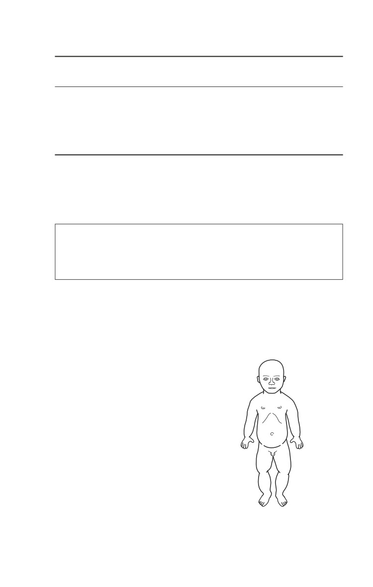
Table 33-3
Age
Weight Resp.
Pulse
BP
(kg)
Rate
(systolic)
Premie
< 3
40-60
130-150
42±10
Term
3
40
120-140
60±10
1-5 yr
~10-20
20-30
100-130
95±30
6 -10 yr
20-32
12-25
75-100
100±15
Adolescent
50
12-18
70
120±20
ο Cardiac stroke volume in children is relatively fixed. Therefore,
bradycardia or relative bradycardia can significantly decrease
cardiac output. Stimulation and oxygen therapy are
corrective for over 90% of significant bradycardias in infants.
Limit peripheral IV access attempts to 2 within 60 seconds
for the child in shock, then immediately proceed to
saphenous vein cutdown or intraosseous (IO) infusion (see
Chapter 8, Vascular Access).
Burns.
ο An infant or child’s head tends to encompass more of the
BSA, with the lower extremities being a smaller percentage.
The palm of the child’s hand can be used to estimate 1%
of total BSA for burn calculations (Fig. 33-1).
Gastrointestinal.
ο Reflux is a common finding,
18%
especially in the newborn period.
This predisposes some children to
Back 13%
difficulty with digestion and
9%
9%
frequent emesis.
Unilateral
18%
ο Children are predisposed to
buttock 2.5%
hypoglycemia due to low
glycogen storage capacity of their
Legs 7.5% on
15% 15%
each side
liver. Full-term infants will tolerate
NPO status for approximately 5
days (with an appropriate D10
solution). Premature infants will
tolerate only 3 days of NPO status Fig. 33-1. BSA percentages
prior to the initiation of TPN.
for infants and children.
33.3
Emergency War Surgery
ο A child’s GI tract is very sensitive to most insults, including
electrolyte abnormalities and systemic illnesses. This can
result in an ileus and manifest as feeding intolerance, and
may precipitate necrotizing enterocolitis (NEC).
ο Gastroenteritis with diarrhea, often associated with fevers,
is also a very common cause of severe dehydration.
Hematology and blood volume.
ο Infants have a physiologic anemia during the first 3-5
months with a hematocrit of 30%-33%.
ο Estimates of blood volume are as follows:
Age
Volume (cc/kg)
Premature
85-100
Term
~80
1-3 mo
75-80
> 3 mo
~70
Renal.
ο Infants and young children have a limited ability to
concentrate urine (max 400-600 mOsm/L) and a fixed
ability to excrete sodium, causing an inability to handle
excess sodium, resulting in hypernatremia if they receive
too much sodium. Premature infants are salt wasters and
full-term infants are salt retainers. It takes 6 years to achieve
normal tubular concentrating ability.
ο Infants can excrete water just like an adult. At 2 weeks of age
the glomerular filtration rate (GFR) is 75% of the eventual
adult rate and reaches maximum capacity at 2 years.
ο Total body water is 80% at 32 weeks, 75% at term, 60%
beyond 1 year.
Thermoregulation.
ο Infants and young children are predisposed to heat loss
and they poorly compensate for wide fluctuations in
ambient temperatures. Children have a higher ratio of
body surface area to mass, and therefore are likely to
become dehydrated earlier than adults when febrile.
ο Reduce exposure and keep infants and children in a
regulated warm environment.
Immune system.
33.4
Pediatric Care
ο Premature infants have incomplete development of their
immune system, causing a 60-fold increased risk of sepsis.
All elective surgery in infants under 30 days of age requires
48 hours of prophylactic antibiotics (with anaerobic coverage
added when appropriate) after the first week of life.
ο Early signs of sepsis can include lethargy, intolerance to
feedings, fever, hypothermia, tachycardia, and irritability
before a rise in the white blood cell count.
Evaluation and Diagnosis
History.
ο Duration/location of pain is important in injury diagnosis.
Over one third of pediatric patients evaluated for
abdominal pain lasting more than a few hours will have
an underlying pathologic condition.
ο Any bilious emesis, especially in the newborn period, may
be a sign of intestinal obstruction, and mandates further
workup.
ο GI bleeding requires immediate attention. The character
(bright red vs melena), quantity, and associated stool
history (ie, diarrhea with infectious source or currant-jelly-
like with intussusception) may provide clues as to the
underlying disorder.
Physical examination.
ο Basic ATLS guidelines should direct the initial assessment
and evaluation for all children involved in traumas. It is
essential to keep the patient warm because children are
much more prone to heat loss than adults.
♦ Modified GCS for children < 4 years old:
Verbal Response
V-Score
Appropriate words/social
5
smile/fixes/follows
Cries, but consolable
4
Persistently irritable
3
Restless, agitated
2
None
1
Radiological studies.
33.5
Emergency War Surgery
ο All bilious vomiting in infants and children should be
evaluated with contrast radiographic imaging. As a general
rule contrast enemas are safer as an initial approach.
♦ Ultrasound is an excellent screening test for the
identification of free abdominal fluid and is also used
in cases of abdominal pain to evaluate appendicitis
(noncompressible appendix with fecolith) and
intussusception (target sign), and others.
♦ Constipation is a common complaint in children that
can be readily diagnosed with plain radiographs and
by history.
Treatment
The treatment algorithm shown below provides the proper
sequence for the rapid sequence intubation (RSI) of the
pediatric patient (Fig. 33-2).
Preoxygenate
Atropine sulfate, 0.1-0.5 mg
Sedation
Midazolam HCl,
Thiopental sodium
0.1 mg/kg (5 mg max)
4-5 mg/kg
Cricoid Pressure
Paralysis
Succinylcholine chloride
Weight < 10 kg: 2mg/kg
Weight > 10 kg: 1 mg/kg
Intubate, Check Tube Position, Release Cricoid Pressure
Fig. 33-2. Rapid sequence intubation for the pediatric patient.
33.6
Pediatric Care
Equipment and Supplies
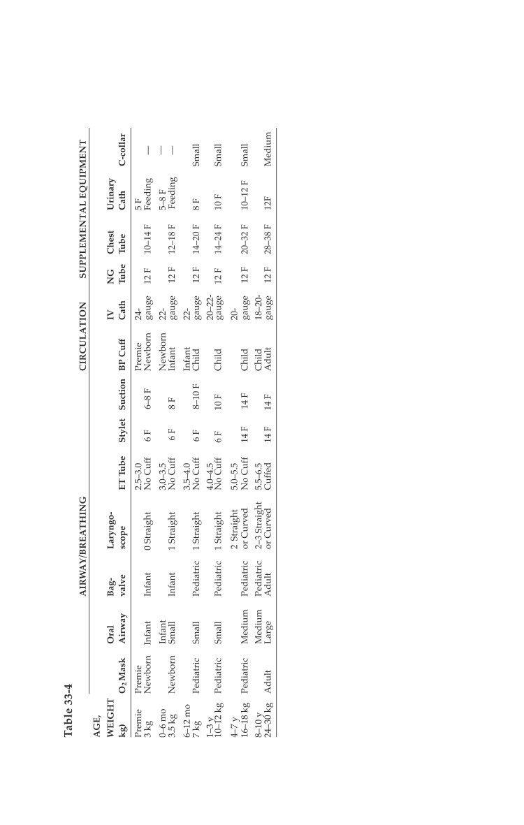
Accessory pediatric medical/surgical equipment arranged
according to age and weight (Table 33-4).
Surgical instruments.
ο If a pediatric surgical set
is not immediately
available, a peripheral
vascular set will usually
contain instruments
delicate enough to
accomplish most tasks in
newborns.
Commonly Used Drugs and
Dosages
Phenobarbital 2-3 mg/kg
IV.
Diazepam 0.25 mg/kg IV
Midazolam HCl 0.1 mg/
kg IV (max 5 mg).
Atropine 0.1-0.5 mg IV.
Phenytoin 15-20 mg/kg,
administered at 0.5 to 1.5
ml/kg/min as a loading
dose, then 4-7 mg/kg/d
for maintenance.
Mannitol 0.5-1.0 g/kg IV.
Succinylcholine chloride 2
mg/kg IV for < 10 kg, and
1 mg/kg IV for > 10 kg.
Ampicillin 25-50 mg/kg IV
q8h (q12-18h in newborns).
Gentamicin 2.5 mg/kg IV
q8h (q12-18h in newborns).
Metronidazole 10 mg/kg
IV.
Acetaminophen 15 mg/kg
po.
33.7
Emergency War Surgery
Surgical Management
Basics.
ο As a general guideline, transverse incisions should be used
in infants. This minimizes the risk of postoperative
dehiscence, while still allowing adequate exposure.
ο Absorbable suture such as Vicryl or PDS (2-0) should be
used to close the rectus fascia, regardless of the incision.
The skin can then be closed using staples or absorbable
monofilament suture (eg, Monocryl 4-0).
ο When placing retention sutures for severe malnutrition,
permanent sutures such as Prolene or nylon can be used
as a full thickness through the rectus muscle and skin. Care
should be taken to avoid the epigastric vessels. The sutures
can be passed through small pieces of a 14F red rubber
catheter prior to tying to avoid excess pressure on the skin.
ο Personnel: Remember that if obstetrics is part of the
mission, pediatric support will be required!
33.8
Chapter 34
Care of Enemy
Prisoners of War/Internees
Introduction
Healthcare personnel of the Armed Forces of the United States
have a responsibility to protect and treat, in the context of a
professional treatment relationship and universal principles of
medical ethics, all detainees in the custody of the Armed Forces.
This includes enemy prisoners of war, retained personnel,
civilian internees, and other detainees. For the purposes of this
chapter, all such personnel are referred to as internees.
It is the policy of the Department of Defense that healthcare
personnel of the Armed Forces and the Department of Defense
should make every effort to comply with “Principles of Medical
Ethics Relevant to the Role of Health Personnel, Particularly
Physicians, in the Protection of Prisoners and Detainees Against
Torture and Other Cruel, Inhuman or Degrading Treatment or
Punishment,” adopted by United Nations General Assembly
Resolution 37/194 of 18 December 1982 (and provided as
Appendix 1 to this book). This is in addition to compliance with
all applicable DoD issuances.
The Geneva Conventions
Define medical personnel as those individuals “exclusively
engaged in the search for, or the collection, transport, or
treatment of the wounded or sick, or in the prevention of
disease; and staff exclusively engaged in the administration
of medical units and establishments” (Geneva Convention
for the Amelioration of the Wounded and Sick in Armed
Forces in the Field [GWS]).
34.1